Pulmonary Department
Mayo Clinic Arizona
Scottsdale, AZ USA
History of Present Illness
The patient is a 73-year-old woman from Wisconsin seen in January 2024 for lung nodules. She had been followed by her physician in Wisconsin for lung nodules but had never had a biopsy or specific diagnosis. She reported that the nodules “waxed and waned.” Her Wisconsin physician suggested she be evaluated in Arizona.
She has occasional cough attributed to paroxysmal nocturnal dyspnea, but denies sputum production, fever, chills or shortness of breath
Past Medical History, Family History and Social History
- Rheumatoid arthritis diagnosed in her 30s, although not currently on any treatment.
- Breast cancer 2006, treated with chemoradiation
- Osteoporosis
- Family history: negative for lung cancer or other lung disorders
- Social History: Lifelong nonsmoker
Medications
- None
Physical Examination
- Unremarkable
Laboratory
- Normal CBC
- Cocci serology: negative
- Rheumatoid factor: elevated 61 U/ml (normal < 15)
- Anti-cyclic citrullinated peptide antibody: negative
- Erythrocyte Sedimentation Rate: normal
Radiology
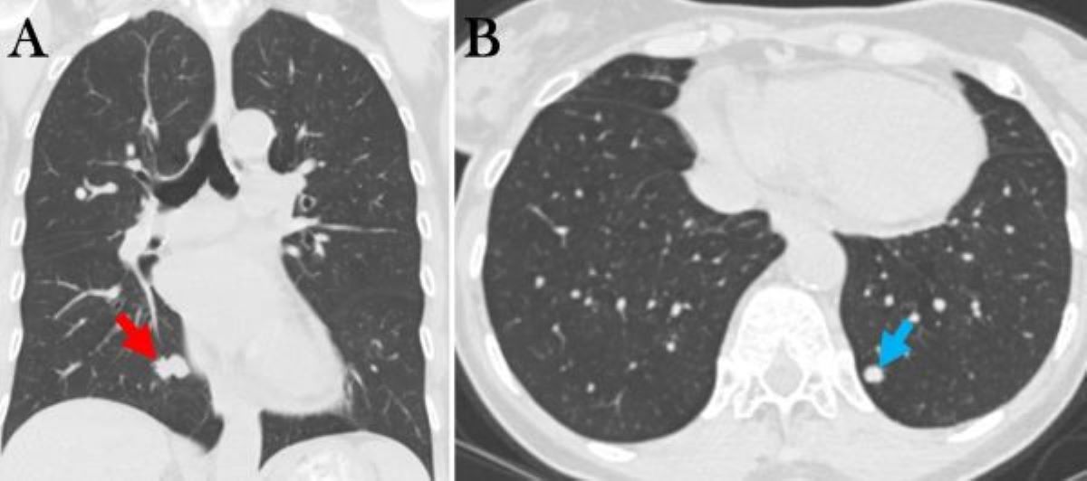
A thoracic CT of the chest done in Wisconsin in November 2023 showed an 18 mm nodule in medial right lower lobe (RLL, Figure 1A) and several other smaller nodules noted, largest other nodule in left lower lobe (LLL, Figure 1B, blue arrow).
 Figure 1. Selected images from thoracic CT done November 2023 showing RLL mass (A, red arrow) and LLL mass (B, blue arrow).
What is the next appropriate step in her evaluation? (Click on the correct answer to be directed to the second of six pages)
- Repeat the thoracic CT scan
- Bronchoscopy
- Positron emission tomography (PET) scan
- 1 and 3
- All of the above