Search Journal-type in search term and press enter
Southwest Pulmonary and Critical Care Fellowships

Arizona Thoracic Society Notes & Videos

(Click on title to be directed to posting, most recent listed first)

January 2020 Video (Passcord TX8x3!%5)
September 2021 Video (Passcode k?6X!z@V)
June 2021 Video (Passcode S1zd7$6g)
December 2020 Arizona Thoracic Society Notes
September 2019 Arizona Thoracic Society Notes
November 2018 Arizona Thorcic Society Notes
September 2018 Arizona Thoracic Society Notes 
July 2018 Arizona Thoracic Society Notes
March 2018 Arizona Thoracic Society Notes
January 2018 Arizona Thoracic Society Notes
November 2017 Arizona Thoracic Society Notes
September 2017 Arizona Thoracic Society Notes
March 2017 Arizona Thoracic Society Notes
January 2017 Arizona Thoracic Society Notes
November 2016 Arizona Thoracic Society Notes
July 2016 Arizona Thoracic Society Notes
March 2016 Arizona Thoracic Society Notes
November 2015 Arizona Thoracic Society Notes
September 2015 Arizona Thoracic Society Notes
July 2015 Arizona Thoracic Society Notes
May 2015 Arizona Thoracic Society Notes
March 2015 Arizona Thoracic Society Notes
January 2015 Arizona Thoracic Society Notes
November 2014 Arizona Thoracic Society Notes
September 2014 Arizona Thoracic Society Notes
August 2014 Arizona Thoracic Society Notes
June 2014 Arizona Thoracic Society Notes
May 2014 Arizona Thoracic Society Notes
April 2014 Arizona Thoracic Society Notes
March 2014 Arizona Thoracic Society Notes
February 2014 Arizona Thoracic Society Notes
January 2014 Arizona Thoracic Society Notes
December 2013 Arizona Thoracic Society Notes
November 2013 Arizona Thoracic Society Notes
October 2013 Arizona Thoracic Society Notes
September 2013 Arizona Thoracic Society Notes
August 2013 Arizona Thoracic Society Notes
July 2013 Arizona Thoracic Society Notes
June 2013 Arizona Thoracic Society Notes
May 2013 Council of Chapter Representatives Notes
May 2013 Arizona Thoracic Society Notes
April 2013 Arizona Thoracic Society Notes 
March 2013 Arizona Thoracic Society Notes
March 2013 Council of Chapter Representatives Meeting 
   and “Hill Day” Notes
February 2013 Arizona Thoracic Society Notes
January 2013 Arizona Thoracic Society Notes
November 2012 Arizona Thoracic Society Notes
October 2012 Arizona Thoracic Society Notes
September 2012 Arizona Thoracic Society Notes
August 2012 Arizona Thoracic Society Notes
August 2012 Special Meeting Arizona Thoracic Society Notes
June 2012 Arizona Thoracic Society Notes
May 2012 Council of Chapter Representatives Meeting

The Arizona Thoracic Society currently has only virtual meetings about 4 times per year. These have been occurring on a Wednesday evening at 7 PM and last until about 8-8:30 PM. 

-------------------------------------------------------------------------------------

Thursday
Nov212013

November 2013 Arizona Thoracic Society Notes

The November Arizona Thoracic Society meeting was held on Wednesday, 11/20/2013 at Shea Hospital beginning at 6:30 PM. There were 26 in attendance representing the pulmonary, critical care, sleep, nursing, radiology, and infectious disease communities.

As per the last meeting a separate area for upcoming meetings has been created in the upper left hand corner of the home page on the SWJPCC website.

A short presentation was made by Timothy Kuberski MD, Chief of Infectious Disease at Maricopa Medical Center, entitled “Clinical Evidence for Coccidioidomycosis as an Etiology for Sarcoidosis”. Isaac Yourison, a medical student at the University of Arizona, will be working with Dr. Kuberski on his scholarly project. Mr. Yourison hypothesizes that certain patients diagnosed with sarcoidosis in Arizona really have coccidioidomycosis. It would be predicted that because of the immunosuppression, usually due to steroids, the sarcoidosis patients would eventually express the Coccidioides infection. The investigators will be collaborating with the University of Washington to perform polymerase chain reaction (PCR) on tissue samples diagnosed with sarcoidosis for Coccidioides.

There were 4 cases presented:

  1. The first case was presented by Lewis Wesselius from the Mayo Clinic Arizona. The patient was a 56 year old woman with rheumatoid arthritis and a prior history of bronchiectasis. In 2009 she was diagnosed with Mycobacterium avium-intracellulare (MAI) on bronchoscopy and started on azithromycin, ethambutol, and rifabutin. She had been on etanercept which was held after her diagnosis of MAI.  She had a negative sputum culture for MAI in September 2012 and her MAI medications were stopped. However, in May 2013 she had increasing symptoms and bronchoscopy demonstrated Pseudomonas and nontuberculous mycobacterium (NTB). She subsequently moved to Phoenix and a CT scan showed the size of her lung nodules to be increased. Bronchoscopic cultures showed Pseudomonas and Mycobacterium abscessus only sensitive to amikacin. She was treated with tigecycline and inhaled amikacin. A repeat CT scan indicated some decrease in size of lung nodules. Dr. Wesselius gave a short presentation on bronchiectasis associated with rheumatoid arthritis and NTB infection in these patients.
  2. The second case was presented by Gerry Swartzberg. Dr. Schwartzberg showed a chest x-ray and asked the audience to guess the diagnosis. Jasminder Mand was the first to correctly guess allergic bronchopulmonary aspergillosis (ABPA) because of the finger in glove sign which best seen in the right upper lobe. The density forms from mucous impaction in a more central bronchus and has been referred to as a rabbit ear appearance, Mickey Mouse appearance, toothpaste shaped opacities, Y-shaped opacities, and V-shaped opacities. Dr. Mand also referred to this as the Churchill sign since it looks like the “V” gesture often associated with Churchill. The patient was begun on corticosteroids and a repeat chest x-ray taken about a month later showed near clearing of the opacities.
  3. Dr. Schwartzberg presented a second case of an elderly woman in her 80’s with a history of bronchiectasis. Chest x-ray and CT scan showed several rapidly expanding lung masses. The radiographic appearance was not particularly suggestive of a diagnosis. There was a concern for malignancy and the majority thought bronchoscopy would be appropriate.
  4. The last case was presented by Joshua  Jewell, a third year pulmonary fellow in the Good Samaritan/VA program. The patient was a middle-aged man who had a history of diffusely metastatic hepatocellular cancer including to his lung and mediastinal lymph nodes. He was also diagnosed with sleep apnea and begun on continuous positive airway pressure (CPAP). He had increasing size of his neck and presented to the pulmonary clinic. Palpation revealed crepitus and a chest x-ray and CT scan confirmed the presence of subcutaneous air and a pneumomediastinum. Dr. Jewell hypothesized that the air was introduced or at least was exacerbated by the CPAP possibly from a ball valve mechanism. Most in the audience agreed this was a reasonable explanation but none had observed this phenomenon previously.

There being no further business the meeting was adjourned at about 8:30 PM. The next meeting is scheduled for Saturday, December 14, 8-12 AM in Tucson at the Kiewit Auditorium at the University of Arizona Medical Center.  The next meeting in Phoenix will be held on Wednesday, January 22, 2014, 6:30 PM at Scottsdale Shea hospital.

Richard A. Robbins, M.D.

Reference as: Robbins RA. November 2013 Arizona thoracic society notes. Southwest J Pulm Crit Care. 2013:7(5):311-2. doi: http://dx.doi.org/10.13175/swjpcc167-13 PDF

Thursday
Oct242013

October 2013 Arizona Thoracic Society Notes

The October Arizona Thoracic Society meeting was held on Wednesday, 10/23/2013 at Shea Hospital beginning at 6:30 PM. There were 21 in attendance representing the pulmonary, critical care, sleep, and thoracic surgery communities.

A proposal was made to decrease the number of meetings from 10 to 8 per year. After a brief discussion, this was adopted. Dr. Parides will try and coordinate these changes with Tucson.

Meetings were announced for December in Tucson, January in Carmel, February in Albuquerque, and April in Phoenix. A suggestion was made to have a separate area for meetings on the SWJPCC website.

There were 2 cases presented-both by Nick Sparacino, a first year fellow at Good Samaritan/VA.  

  1. The first case was a 48 year old man admitted to podiatry for chronic diabetic foot ulcers.  His preoperative chest x-ray revealed multiple pulmonary nodules. Importantly, he had a history of working in a brake pad factory for about 15 years, a strong family history of lung cancer and was currently actively smoking. Review of the chest x-ray and the CT scan revealed that pleural nodules only on the left. Additional history was obtained of a gunshot wound through the spleen into the chest. A liver-spleen scan showed high uptake in the nodules. The nodules were thought to be secondary to thoracic splenosis which occurs when splenic tissue is autoimplantated to the thoracic cavity following splenic injury (1). No further work up or therapy was thought to be needed.
  2. The second case was a 66 year old man with 2-3 week history of shortness of breath, subjective fevers, sputum production, two falls without injury, and urinary incontinence. Chest x-ray showed right lower lobe pneumonia and CT scan of the chest showed narrowing of the bronchus intermedius. Bronchoscopy revealed a veruccous, obstructing mass in the bronchus intermedius that was suspicious for squamous cell carcinoma. However, on biopsy the mass separated from the bronchial wall and fractured. It was eventually removed piecemeal with the flexible fiberoptic bronchoscope (1). Pathology was consistent with a walnut.

There being no further business the meeting was adjourned at about 8 PM. The next meeting is scheduled for Wednesday, November 20, 6:30 PM in Phoenix at Scottsdale Shea Hospital. 

Richard A. Robbins, M.D.

References

  1. Khan AM, Manzoor K, Gordon D, Berman A. Thoracic splenosis: A diagnosis by history and imaging. Respirology. 2008;13(3):481-3. [CrossRef] [PubMed]
  2. Boyd M, Chatterjee A, Chiles C, Chin R Jr. Tracheobronchial foreign body aspiration in adults. South Med J. 2009;102(2):171-4. [CrossRef]

Reference as: Robbins RA. October 2013 Arizona thoracic society notes. Southwest J Pulm Crit Care. 2013;7(4):253-4. doi: http://dx.doi.org/10.13175/swjpcc144-13 PDF

Saturday
Sep282013

September 2013 Arizona Thoracic Society Notes

The September Arizona Thoracic Society meeting was held on Wednesday, 9/25/2013 at Shea Hospital beginning at 6:30 PM. There were 13 in attendance representing the pulmonary, critical care, sleep, and pathology communities.

After a brief discussion, Gerry Swartzberg was selected as Arizona’s 2014 nominee for Clinician of the Year. 

There was 1 case presented:

Dr. Thomas Colby, pulmonary pathologist from Mayo Clinic Arizona, presented the case of a 67 year old woman with multiple pulmonary nodules. The largest was 1.2 cm CT scan. She had a fine needle aspiration of one of the nodules. The pathology revealed spindle-shaped cells which were synaptophysin + (also known as the major synaptic vesicle protein p38). Synaptophysin marks neuroendocrine tissue and on this basis the patient was diagnosed with multiple carcinoid tumors. Aguayo et al. (1) described six patients with diffuse hyperplasia and dysplasia of pulmonary neuroendocrine cells, multiple carcinoid tumorlets, and peribronchiolar fibrosis obliterating small airways. Miller and Müller (2) described a series of 25 patients that were mostly women. Eight had obliterative bronchiolitis. Many questions arose including PET positivity (variable), endobronchial spread (unknown), use of somatostatin receptor scintigraphy (unknown).

There being no further business the meeting was adjourned at about 7:30 PM. The next meeting is scheduled for Wednesday, October 23, 6:30 PM in Phoenix at Scottsdale Shea Hospital.  

Richard A. Robbins, M.D.

References

  1. Aguayo SM, Miller YE, Waldron JA Jr, Bogin RM, Sunday ME, Staton GW Jr, Beam WR, King TE Jr. Brief report: idiopathic diffuse hyperplasia of pulmonary neuroendocrine cells and airways disease. N Engl J Med. 1992;327(18):1285-8. [CrossRef] [PubMed]
  2. Miller RR, Müller NL. Neuroendocrine cell hyperplasia and obliterative bronchiolitis in patients with peripheral carcinoid tumors. Am J Surg Pathol. 1995;19(6):653-8. [CrossRef] [PubMed]

Reference as: Robbins RA. September 2013 Arizona thoracic society notes. Southwest J Pulm Crit Care. 2013;7(3):205. doi: http://dx.doi.org/10.13175/swjpcc132-13 PDF

Thursday
Aug292013

August 2013 Arizona Thoracic Society Notes

The August Arizona Thoracic Society meeting was held on Wednesday, 8/28/2013 at Shea Hospital beginning at 6:30 PM. There were 23 in attendance representing the pulmonary, critical care, sleep, and pathology communities.

A brief discussion was held about the audio-visual aids available. It was generally agreed that our current projector is inadequate. Judd Tillinghast will inquire about using a hospital overhead projector. If that is not possible, it was agreed to purchase a new projector.Plans for telecasting the meeting between Phoenix and Tucson continue. A trial of a link between Shea and the University in Tucson failed. Once the link is successfully established, it is hoped that the meeting can be telecasted.

There were 6 cases presented:

1. Dr. Thomas Colby, pulmonary pathologist from Mayo Clinic Arizona, presented the case of a 10 year old boy with chronic dyspnea for > 4 yrs. He had growth retardation since age 1, a skin rash since age 2 on the limbs, nail dystrophy since age 3 on hands and feet, lacrimal duct stenosis, erythematous lesions on the pinnae, phimosis, interstitial lung disease on radiography, weakly positive p-ANCA, elevated erythrocyte sedimentation rate, and hypergammaglobulinemia. He came to lung biopsy. The patient was diagnosed with dyskeratosis congenita which is a disorder of poor telomere maintenance (1). Specifically, the disease is related to one or more mutations which directly or indirectly affect the vertebrate telomerase RNA component (TERC). This patient’s manifestations are fairly typical of the disease. Short telomere length was confirmed.

2. Dr. Colby presented a second case of a 14 year old boy with a history of osteosarcoma. Pulmonary nodules developed and biopsy showed metastatic osteosarcoma. He was given systemic chemotherapy but now has residual nodules that were biopsied. One of the pulmonary nodule resembled bronchoalveolar cell carcinoma.  This is an apparent complication following chemotherapy in adolescent patients (2).

3. Dr. Colby presented the pathology of a patient from the Phoenix VA who underwent lung biopsy for interstitial disease. The pathology was typical for IgG4-related disease with a plasma cell rich lymphohistiocytic infiltrate in the bronchovascular sheath and histopathology showing diffusely stained positive for IgG4 plasma cells (3).

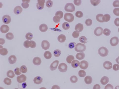
4. Dr. Suresh Uppalapu, a second year pulmonary fellow from Good Samaritan/VA, presented a 59 year old Sudanese male who was transferred to the Good Samaritan ICU in shock. His presenting complaints to the transferring hospital were acute mental status changes, weakness, and chills. He was intubated for hypercarbic respiratory failure. His brother related that the patient had just returned from Sudan three weeks earlier. He had a prior history of a splenectomy. He was hypothermic with a temperature of 32.3°C and a SpO2 of 91% on 100% FiO2 and PEEP of 8. His Glasgow Coma Scale was 3 (lowest possible score). He had many abnormalities on laboratory evaluation, most notably a creatinine of 5.1 mg/dL and a lactic acid of 26.3 mg/dL. The peripheral smear showed malaria parasites typical of falciparum malaria (figure 1).

Figure 1. Peripheral smear showing a gametocyte (red arrow) and trophozoites in various stages from falciparum malaria.

He developed hemoptysis and eventually expired. A preliminary autopsy report has detected aspergillosis in the lung. Invasive aspergillosis has been reported in cases of severe falciparum malaria (4).

5. Dr. Heemesh Seth, also a second year pulmonary fellow from Good Samaritan/VA, presented a case of a 57 year old man with cirrhosis secondary to hepatitis C diagnosed in 1998. He presented with a large right hydrothorax.  Multiple thoracentesis were performed (Table 1).

Table 1. Summary of multiple thoracentesis.

Blood cultures were positive for acinetobacter as was the initial culture from the thoracentesis. He was treated with cephepime. It was felt that his effusion and empyema were most likely secondary to translocation of bacteria to the pleural space from spontaneous bacterial peritonitis. A discussion ensued regarding whether to perform tube thoracostomy. Data is sparse with most literature not favoring a chest tube (5). However, in this patient’s case a chest tube was eventually inserted when he failed to improve. It drained about 2 liters of fluid but the drainage then became minimal and the tube was removed. The patient developed hepato-renal syndrome but was felt not to be a liver transplant candidate. He was transferred to hospice.

6. Dr. Seth also presented a second case of a 66 year old Hispanic man who presented with a large left pleural effusion.  He had a past medical history of systemic lupus erythematosis (SLE) with possible rheumatoid arthritis and was being treated with adalimumab, methotrexate, and prednisone. A thoracentesis was done and 2 liters of clear amber fluid was removed. Although be developed fever to 102°F he felt much better the next morning and was discharged. However, his coccidioidomycosis serologies were positive for both IgG and IgM and his complement fixation test were positive at 1:4. Pleural fluid cytology was positive for LE cells. He was continued on prednisone and treated with fluconazole. A discussion developed of whether the effusion was secondary to SLE, coccidioidomycosis, or both. No one knew data but it was felt that it was most prudent to continue the present course while following the patient and awaiting cultures.

There being no further business the meeting was adjourned at about 8:15 PM. The next meeting is scheduled for Wednesday, September 25, 6:30 PM in Phoenix at Scottsdale Shea Hospital.  

Richard A. Robbins, M.D.

References

  1. Dokal I. Dyskeratosis congenita in all its forms. Br J Haematol. 2000;110(4):768-79. [CrossRef] [PubMed] 
  2. Travis WD, Linnoila RI, Horowitz M, Becker RL Jr, Pass H, Ozols R, Gazdar A. Pulmonary nodules resembling bronchioloalveolar carcinoma in adolescent cancer patients. Mod Pathol. 1988;1(5):372-7. [PubMed]
  3. Hurley JR, Leslie KO. IgG4-Related systemic disease of the pancreas with involvement of the lung: a case report and literature review. Southwest J Pulm Crit Care. 2013;7(2):117-130. [CrossRef]
  4. Hocqueloux L, Bruneel F, Pages CL, Vachon F. Fatal invasive aspergillosis complicating severe Plasmodium falciparum malaria. Clin Infect Dis. 2000;30(6):940-2. [CrossRef] [PubMed] 
  5. Alonso JC. Pleural effusion in liver disease. Semin Respir Crit Care Med. 2010;31(6):698-705. [CrossRef] [PubMed] 

Reference as: Robbins RA. August 2013 Arizona Thoracic Society notes. Southwest J Pulm Crit Care. 2013;7(2):114-6. doi: http://dx.doi.org/10.13175/swjpcc117-13 PDF

Thursday
Jul252013

July 2013 Arizona Thoracic Society Notes

The first Arizona Thoracic Society meeting in Tucson was held on Wednesday, 7/24/2013 at Kiewit Auditorium on the University of Arizona Medical Center campus beginning at 6:30 PM. There were 36 in attendance representing the pulmonary, critical care, sleep, pathology and radiology communities. Dinner was sponsored by Accredo Health Group.

A brief discussion was held of plans to have the December 2013 meeting in Tucson on a weekend as part of the University of Arizona winter pulmonary meeting.

There were 4 cases presented:

  1. Mohammad Dalabih presented a case of a 48 yo woman with respiratory failure cared for by Gordon Carr, Linda Snyder, and himself. Radiology findings were discussed by Isabel Oliva. Lung biopsy showed acute fibrinous and organizing pneumonia rather than ARDS and was presented by Richard Sobonya.
  2. Franz Rischard presented a case of a 61 year old with progressive dyspnea and moderate COPD with evidence of pulmonary hypertension. Radiologic work up was presented by Dr. Oliva. It was determined the patient had primary hypertension and Dr. Rischard discussed how to separate pulmonary hypertension from COPD from primary pulmonary hypertension.
  3. Nathaniel Reyes presented a 50 year old woman with a history of granulomatous polyangiitis (GPA, formerly known as Wegener’s granulomatosis) who was pANCA+ but cANCA-. She developed diffuse alveolar hemorrhage. Dr. Soboyna reviewed pathology of GPA. Dr. Reyes discussed the ANCA+ vasculities. Some rheumatologists no longer consider GPA and microscopic polyangitis separate diseases but part of the same spectrum.
  4. Gordon Carr presented a case of a 65 year old man who died of an exacerbation of idiopathic pulmonary fibrosis (IPF). Dr. Sobonya reviewed the autopsy and pathology findings of IPF.

There being no further business the meeting was adjourned at about 8:15 PM. The next meeting is scheduled for Wednesday, August 28, 6:30 PM in Phoenix at Scottsdale Shea Hospital.  

Richard A. Robbins, M.D.

Reference as: Robbins RA. July 2013 Arizona thoracic society notes. Southwest J Pulm Crit Care. 2013;7(1):50. doi: http://dx.doi.org/10.13175/swjpcc095-13. PDF 

Page 1 ... 5 6 7 8 9 ... 14 Next 5 Entries »