Search Journal-type in search term and press enter
Southwest Pulmonary and Critical Care Fellowships

Critical Care Journal Club

(Click on title to be directed to posting, most recent listed first)

November 2017 Phoenix Critical Care Journal Club
Tucson Critical Care Journal Club: Albumin Use in the Critical Care Unit
May 2016 Phoenix Critical Care Journal Club: Oxygen Therapy
November 2015 Tucson Critical Care Journal Club: Atrial Fibrillation in
   Sepsis
September 2015 Phoenix Critical Care Journal Club: Goal-Directed
Therapy and DNR
June 2015 Phoenix Critical Care Journal Club: Interventions in ARDS
February 2015 Phoenix Critical Care Journal Club: Intracranial Pressure
Monitoring for Fulminant Liver Failure
August 2014 Tucson Critical Care Journal Club: Bacteremia in Cardiac 
   Arrest
July 2014 Phoenix Critical Care Journal Club: Predicting the Future
April 2014 Tucson Critical Care Journal Club: Early Goal-Directed
   Therapy
April 2014 Phoenix Critical Care Journal Club: Early Goal-Directed
   Therapy
March Critical Care Journal Club: Sequelae of Critical Care
February 2014 Phoenix Critical Care Journal Club: Subgroup Analysis
January 2014 Critical Care Journal Club
November 2013 Critical Care Journal Club
September 2013 Banner Good Samaritan / Phoenix VA Critical Care
   Journal Club
September 2013 Tucson Critical Care Journal Club: Early
   Tracheostomy
August 2013 Critical Care Journal Club: Less is More
July 2013 Critical Care Journal Club
April 2013 Critical Care Journal Club
March 2013 Critical Care Journal Club
February 2013 Critical Care Journal Club
January 2013 Critical Care Journal Club
December 2012 Critical Care Journal Club
November 2012 Critical Care Journal Club
October 2012 Critical Care Journal Club
September 2012 Critical Care Journal Club
August 2012 Critical Care Journal Club
July 2012 Critical Care Journal Club
June 2012 Critical Care Journal Club
May 2012 Critical Care Journal Club
April 2012 Critical Care Journal Club
February 2012 Critical Care Journal Club
January 2012 Critical Care Journal Club
December 2011 Critical Care Journal Club 
November 2011 Critical Care Journal Club
September 2011 Critical Care Journal Club 
August 2011 Critical Care Journal Club 
July 2011 Critical Care Journal Club
June 2011 Critical Care Journal Club 
April 2011 Critical Care Journal Club
March 2011 Critical Care Journal Club
February 2011 Critical Care Journal Club
November 2010 Critical Care Journal Club
October 2010 Critical Care Journal Club


Both the Phoenix Good Samaritan/VA and the University of Arizona fellows previously had a periodic critical care journal club in which current or classic articles were reviewed and discussed. A brief summary was written of each discussion describing the article and the strengths and weaknesses of each article.

-------------------------------------------------------------------------------------

Friday
Apr262013

April 2013 Critical Care Journal Club

We welcomed intensivists from Banner Health to video-conference with us as we discussed several articles, and evaluated the ACP Journal Club – another good resource for keeping up to date.

Hill NS. Review: Lower rather than higher tidal volume benefits ventilated patients without ARDS. Ann Intern Med. 2013;158:JC4. Abstract

Lauzier F. Hydroxyethyl starch 130/0.4 and saline did not differ for mortality at 90 days in ICU patients. Ann Intern Med. 2013;158:JC5. Abstract

The April ACP Journal Club reviewed two critical care articles – a meta-analysis that concluded that low tidal volume ventilation reduced mortality in patients without ARDS, and a large RCT that showed no mortality difference between critically-ill patients resuscitated with hydroxyethyl starch versus saline.  Both articles were awarded 6/7 stars for “clinical impact”, yet neither article had any impact on our clinical practice.  This troubled us.

We could think of 4 necessary criteria in order for research to have legitimate clinical impact: 1) internal validity; 2) external applicability; 3) an intervention that is different from what we are already doing; and 4) a clinically significant benefit.  We had previously critically appraised the study on low tidal volume ventilation in non-ARDS patients, found it sorely lacking in internal validity, therefore rejecting the practice for clinical application.  Two-thirds of the patients included in this meta-analysis were enrolled in studies that were only observational in design.  Many of the included studies focused on short duration, post-op mechanical ventilation.  It seemed biologically implausible to us that alveolar hyperinflation could lead to ventilator-associated lung injury in patients intubated for a few days with essentially normal post-op lungs.  The ACP reviewer endorsed low tidal volume ventilation in non-ARDS patients in part because of the “lack of evidence to suggest harm”.  Yet low tidal ventilation can be uncomfortable for an awake patient, and can only be maintained in some by increasing sedation medications.  There is good evidence that this can lead to prolonged ventilator LOS and associated complications.  Observational studies lumped into a meta-analysis should only be used to generate hypotheses.  Therefore, we would have rated this article as 0/7 stars for clinical impact - no change warranted in clinical practice, but further study recommended.

The second critical care study in the ACP journal club has high internal validity, but did not change our practice either, because we already knew from multiple previous studies that colloid resuscitation fluids were not superior to crystalloids, and increase the risk of acute kidney injury in critically-ill patients.  We would have rated this article as 1/7 stars for clinical impact because it might change the practice of a minority of clinicians who are still routinely using colloid resuscitation, if the previous studies hadn’t already convinced them to do so. 

It is our observation that the reviewers for ACP journal club generally overestimate the clinical impact of the studies they review.  We recommend that they should develop a precise and reasonable definition of “clinical impact” such as we have proposed, and use it to provide more accurate critical appraisal ratings.   

Ioannidis JP. Are mortality differences detected by administrative data reliable and actionable? JAMA. 2013;309:1410-1. Abstract

We have frequently reviewed articles that use DRG billing codes databases.  The convenience and large size of these databases makes them tempting to analyze, but it has been repeatedly demonstrated that they are often not accurate for research purposes.  Financial incentives have been shown to significantly alter hospital coding procedures resulting in over or under-coding of certain diagnoses, comorbidities and complications (depending on the incentives).  Billing codes that don’t accurately reflect the patient’s true clinical condition can lead to errors in identifying patients with the diagnosis of interest, adjusting for severity of illness, and determining outcomes and interventions might have affected the outcome.  Administrative database studies may also be overpowered.  This might seem like a good thing, but very large sample sizes can result in “statistically significant” differences that are clinically infinitesimally small.  We feel that observational studies based on administrative databases should be viewed as hypothesis-generating.  In almost all cases, their internal validity is inferior to observational studies based on clinical data.

Weber-Carstens S, Schneider J, Wollersheim T, Assmann A, Bierbrauer J, Marg A, Al Hasani H, Chadt A, Wenzel K, Koch S, Fielitz J, Kleber C, Faust K, Mai K, Spies CD, Luft FC, Boschmann M, Spranger J, Spuler S. Critical illness myopathy and GLUT4: significance of insulin and muscle contraction. Am J Respir Crit Care Med. 2013;187:387-96. Abstract

This study sought to clarify the mechanism of critical illness myopathy, and test the concept of whether electrically-induced muscle stimulation might prevent it.  It was not meant to immediately impact clinical care, but to guide further research.  I appreciate Emad volunteering to review it, as it is very intimidating reading.  I’m going to condense our take on it.

The authors screened 874 critically ill patients in order to complete a study of only 35.  Thirty of these underwent electrophysiological testing with an insulin clamp, of which 12 had critical illness myopathy (CIM), and 18 did not.  The other five patients underwent electrical stimulation of a unilateral vastus lateralis muscle in an attempt to prevent CIM, using the contralateral unstimulated muscle as a control. 

Normally, glucose uptake in muscle cells is facilitated by glucose transporter 4  (GLU4) – which is stored in perinuclear vesicles, and rapidly released and translocated to the sarcolemma in response to insulin receptor binding, or muscle contraction.   However, it was found that this mechanism fails in critical illness.  Muscle glucose utilization was not stimulated by high-dose insulin in study subjects.  Insulin resistance was demonstrated in all the critically-ill patients, but was worse in those with CIM, in whom immunohistochemical studies of muscle tissue showed that GLU4 was essentially trapped in the perinuclear vesicles.  Several signal transduction proteins that regulate GLU4 were shown to be significantly under-expressed in these patients – perhaps explaining why this might be. 

In the patients who underwent periodic electrical muscle stimulation during their illness, these transduction proteins were restored, GLU4 release into the sarcolemma was maintained, and the muscle fibers showed robust morphological improvements compared to the unstimulated contralateral leg muscles.  This study helps expand our understanding of the pathogenesis of critical illness myopathy and insulin resistance, and suggests a method to prevent CIM that deserves further clinical study. 

Prasad V, Rho J, Cifu A. The inferior vena cava filter: how could a medical device be so well accepted without any evidence of efficacy? JAMA Intern Med. 2013;173:493-5. Abstract

Although the inferior vena cava (IVC) filter is FDA-approved, and use has steadily climbed to over 50,000 device placements per year, there is no valid evidence of clinical efficacy.  The study that initially supported use of IVC filters was an uncontrolled case series reported by Greenfield.  He observed a 4% rate of pulmonary embolism (PE) after IVC filter placement, but nearly half the patients were lost to follow-up.  The true rate of PE in patients with Greenfield filters is unknowable in such a study, because it’s possible that patients who suffered PE were more likely to be lost to follow-up.  A single randomized controlled trial (RCT) addresses the clinical efficacy of IVC filters.  Four-hundred patients with venous thromboembolism were randomized to receive IVC filters in addition to antithrombotic therapy.  Interestingly, patients who had the most widely accepted indication of IVC filters – those with contraindications to antithrombotic therapy - were excluded from this study.  At a two-year follow-up, there was no significant reduction in PE or mortality in the group with IVC filters, but a statistically significant absolute increase in the rate of deep venous thrombosis by 10%.  At an eight-year follow-up, the rate of PE as elicited by an annual phone interview was statistically significantly reduced in patients with an IVC filter.  The rate of DVT remained significantly higher in patients with filters, and mortality remained unaffected. 

This RCT has little clinical relevance since IVC filters are not usually recommended for the type of patients enrolled in the study.  Nor are IVC filters placed in order to prevent pulmonary embolism occurring more than 2 years later.  The study did show convincingly that IVC filters increase the rate of DVT.  Other studies have shown complications including fracture and fragment embolization, and whole filter embolization which have in some cases caused cardiac arrest and fatality.  One study showed a 33% risk for IVC occlusion at 9 years.  Retrievable filters seemed to have been widely clinically accepted, but retrieving them would mitigate the only possibly proven benefit (preventing PE occurring years later) – in any case, another study suggests that as few as 10% of retrievable filters are ever retrieved. 

The authors challenge the lack of support for FDA approval of these devices, and point out that there is no financial incentive for further research by the manufacturers, so long as physicians are willing to use them without evidence of efficacy. 

A spirited discussion expressed a wide spectrum of views.  We discussed the common use of prophylactic IVC filters in patients with a high risk for primary or recurrent venous thromboembolism – for instance in trauma or cancer patients.  Some felt that the standard of care demands use of IVC filters in this situation, even in the absence of scientific evidence of benefit.  Others felt that IVC filters are highly over-utilized in these situations.  In light of our earlier discussion, this is an article that I feel has legitimate clinical impact.  Many of us will still probably consider IVC filters for patients with acute venous thromboembolism who have strong contraindications to antithrombotic therapy, despite the absence of evidence that it will prevent immediate life-threatening pulmonary embolism.  But this article will likely make us more cautious in recommending this potentially dangerous device to patients with other proposed indications.

In addition to inducing DVT, IVC filters can induce propagation of the clot. If there are contraindications to anticoagulation, this might induce the placement of additional filters, further propagation of the clot, additional filters, etc. (Figure 1).
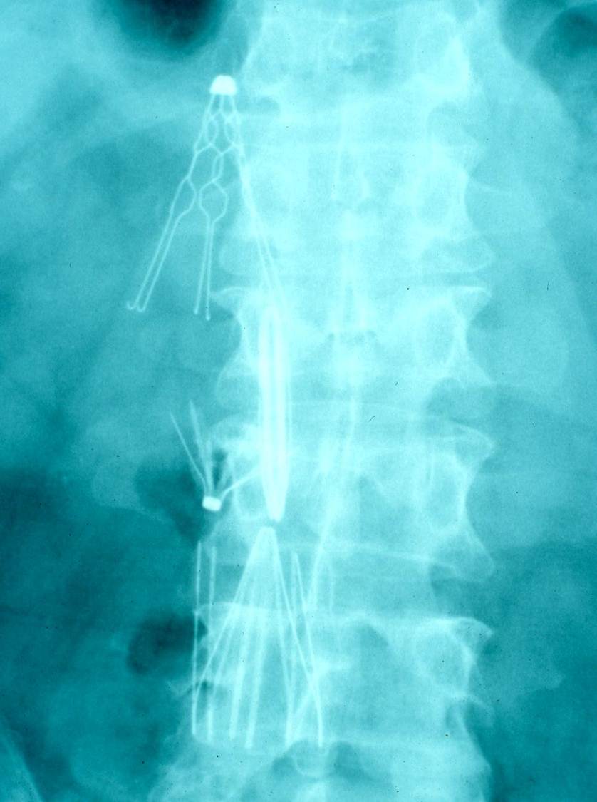
Figure 1. Patient with multiple IVC filters placed.

I was fascinated by our group reaction to this article.  It is apparent that most of us still struggle when our logic conflicts with empirical evidence.  In 1984, Goodwin coined the term “tomato effect” to describe the situation in which an efficacious treatment for a certain disease is ignored or rejected because it doesn’t make sense in light of accepted theories of disease mechanisms.  [The term was chosen because a widely-held misconception in North American in the 1600 and 1700’s that tomatoes were poisonous – causing consumption of tomatoes to lag far behind their acceptance in  Europe].  Goodwin contrasted the tomato effect with the placebo effect.  But in the case of IVC filters, I’m not sure the term placebo effect is applicable.  Many physicians are so convinced that IVC filters ought to work, that they are willing to forego scientific proof that they do work.  Maybe we should call this the “Swan effect” since for years we placed Swan-Ganz pulmonary artery catheters before evidence was published that they provide little or no benefit to the patient.

Robert A. Raschke MD, MS

Associate Editor

Reference as: Raschke RA. April 2013 critical care journal club. Southwest J Pulm Crit Care. 2013;6(4):191-5. PDF

Tuesday
Apr022013

March 2013 Critical Care Journal Club

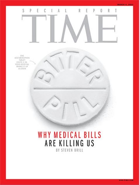
Brill S. Bitter Pill: Why Medical Bills Are Killing Us. Time. February 20, 2013. PDF available at: http://livingwithmcl.com/BitterPill.pdf (accessed 4/2/13).

Editor’s Note: We had a special journal club in March. First, we reviewed an article from Time magazine rather than a traditional medical journal. Second, Paul Stander MD, the chief medical officer at Banner Good Samaritan Regional Medical Center, led the discussion and agreed to author the journal club. This seemed appropriate since much of the article focuses on overbilling and administrative costs of care.

The recent lengthy cover story article in Time Magazine described in great deal what many of us practicing physicians have realized for a long time – our health care system is highly dysfunctional and much of that dysfunction is a result of an arcane and outmoded financing mechanism. This payment system has a litany of perverse incentives that encourage wasteful and often ineffective care while not compensating for necessary, but less glamorous services, while simultaneously doing so at exorbitant cost that has created for us a competitive disadvantage in the world marketplace, contributed mightily to our federal budget deficit and led thousands of hardworking citizens to financial hardship or bankruptcy.  The major points of the article were the following:

  1. Hospital pricing and charging mechanisms have no real relationship to true costs.  This system is a result of decades long price increases that began in the era when hospitals were paid as a percentage of their charges.  These charges for the most part do not currently reflect what hospitals are compensated by insurance companies or the government who pay fixed amounts based on case or per diem rates.  Unfortunately the only people who may pay these inflated prices are those who are least capable of doing so - uninsured individuals who can then be financially devastated.
  2. Pharmaceutical and device manufacturers are paid far higher fees for their products in the United States than anywhere else in the world for no real justifiable reason which contributes to the disparate cost of care in this country compared to others
  3. Not-for-profit hospitals often make very high profit margins and have highly paid executives raising serious questions about the fairness of their tax exempt status and whether they truly fulfill their supposed community mission and whether they are truly providing adequate care for indigent and uninsured populations.
  4. The Affordable Care Act only partially addresses some of these issues – it really is designed to bring a lot more people into this-pro dysfunctional system, many of whom will be covered by government payments.  While this will protect more patients from financial ruin, but it does little to address the other issues described above.

Real reform to our healthcare system will come only when we change the focus of the care we provide – particularly on the growing number of elderly patients with multiple chronic conditions who are the drivers of our ever increasing costs for Medicare and we also change the perverse incentives in our system that encourage waste and spending on ineffective or even harmful interventions.  These reforms must be driven by clinicians in order for them to succeed.

Paul Stander MD

Chief Medical Officer

Banner Good Samaritan Regional Medical Center

Reference as: Stander P. March 2013 critical care journal club. Southwest J Pulm Crit Care. 2013;6(4):168-9. PDF

Wednesday
Feb272013

February 2013 Critical Care Journal Club

Ferguson ND, Cook DJ, Guyatt GH, Mehta S, Hand L, Austin P, Zhou Q, Matte A, Walter SD, Lamontagne F, Granton JT, Arabi YM, Arroliga AC, Stewart TE, Slutsky AS, Meade MO; the OSCILLATE Trial Investigators and the Canadian Critical Care Trials Group. High-Frequency Oscillation in Early Acute Respiratory Distress Syndrome. N Engl J Med. 2013;368:795-805. Abstract

Young D, Lamb SE, Shah S, Mackenzie I, Tunnicliffe W, Lall R, Rowan K, Cuthbertson BH; the OSCAR Study Group. High-Frequency Oscillation for Acute Respiratory Distress Syndrome. N Engl J Med. 2013;368:806-13. Abstract

Malhotra A, Drazen JM. High-Frequency Oscillatory Ventilation on Shaky Ground. N Engl J Med. 2013;368:863-5. Abstract

Two articles and an accompanying editorial, the later co-authored by none less than the editor, appeared in the New England Journal of Medicine this week. These all dealt with the use of high-frequency oscillatory ventilation (HFOV) in the adult respiratory distress syndrome (ARDS). As the editorial points out, HFOV should prove to be advantageous in ARDS. Conventional mechanical ventilation can perpetuate lung injury in ARDS by at least two mechanisms:

1. Excessive tidal volumes can stretch the lung, leading to overdistention and further lung injury.

2. Inadequate positive end-expiratory pressure (PEEP) can promote repetitive alveolar collapse followed by reopening, which may be injurious to the lung (an injury known as atelectrauma).

Theoretically, HFOV should lessen both types of injury since it vibrates gas in the airway resulting in alveolar ventilation.

However, the results of two major, multicenter, randomized trials found no clinical advantage of HFOV. In fact, in one of the trials, the Oscillation for Acute Respiratory Distress Syndrome Treated Early (OSCILLATE) trial, patients treated with HFOV had more deaths than conventional mechanical-ventilation. In the other trial, the OSCAR trial, no difference in 30-day mortality was observed.

The mechanism underlying the failure of HFOV to improve outcomes is unknown although a number of possibilities are discussed in the editorial. Also pointed out is that since ARDS is a heterogenous lung disease from differing causes, some patient subgroups might be helped while others are harmed by HFOV. Regardless, for now clinicians should be cautious about applying HFOV routinely in patients with ARDS.

Richard A. Robbins, MD

Editor, Southwest Journal of Pulmonary and Critical Care

Reference as: Robbins RA. February 2013 critical care journal club. Southwest J Pulm Crit Care 2013;6(2):89. PDF

Tuesday
Jan292013

January 2013 Critical Care Journal Club

Villanueva C, Colomo A, Bosch A, Concepción M, Hernandez-Gea V, Aracil C, Graupera I, Poca M, Alvarez-Urturi C, Gordillo J, Guarner-Argente C, Santaló M, Muñiz E, Guarner C. Transfusion strategies for acute upper gastrointestinal bleeding. N Engl J Med. 2013;368(1):11-21. Abstract

The hemoglobin threshold for transfusion of red cells in patients with acute gastrointestinal bleeding is controversial. The authors compared the efficacy and safety of a restrictive transfusion strategy (transfusion when the hemoglobin level fell below 7 g per deciliter) with a liberal transfusion strategy in 921 patients with severe acute upper gastrointestinal bleeding. The probability of survival at 6 weeks was higher in the restrictive-strategy group than in the liberal-strategy group and the probability of further bleeding adverse events were lower.

This article may alter practices in the ICU. Previously, many intensivists, including myself, thought it was important to make sure that we “stayed ahead” of the bleeding. This study suggests this is a flawed strategy leading to increased bleeding, more complications and increased mortality. Furthermore, the study is consistent with other studies, particularly those where the bleeding results from esophageal varices. In the absence of conflicting data, use of a restrictive strategy is clinically indicated because it significantly improves patient outcomes in patients with acute upper gastrointestinal bleeding.

Chesnut RM, Temkin N, Carney N, Dikmen S, Rondina C, Videtta W, Petroni G, Lujan S, Pridgeon J, Barber J, Machamer J, Chaddock K, Celix JM, Cherner M, Hendrix T. A trial of intracranial-pressure monitoring in traumatic brain injury. N Engl J Med. 2012;367(26):2471-81. Abstract

Intracranial-pressure monitoring is considered the standard of care for severe traumatic brain injury (TBI) and is used frequently, but the efficacy of treatment based on monitoring in improving patient outcomes has not been rigorously assessed. The authors conducted a multicenter, controlled trial of 324 patients with severe TBI. The patients were randomly assigned to one of two specific protocols: guidelines-based management in which a protocol for monitoring intraparenchymal intracranial pressure was used; or a protocol in which treatment was based on imaging and clinical examination. The primary outcome was a composite of survival time, impaired consciousness, and functional status at 3 months and 6 months and neuropsychological status at 6 months. There was no change in the primary outcome and the distribution of serious adverse events was similar in the two groups. For patients with severe TBI, care focused on maintaining monitored intracranial pressure was not shown in this article to be superior to care based on imaging and clinical examination.

Like the restrictive transfusion strategy in upper gastrointestinal bleeds discussed above, this is a good randomized study. Although the study does not show that monitoring is harmful, it does demonstrate that it is ineffective in patients with TBI. In the absence of conflicting data, use of intracranial-pressure monitoring is not clinically indicated in patients with TBI because it does improve patient outcomes.

Sinuff T, Muscedere J, Cook DJ, Dodek PM, Anderson W, Keenan SP, Wood G, Tan R, Haupt MT, Miletin M, Bouali R, Jiang X, Day AG, Overvelde J, Heyland DK; Canadian Critical Care Trials Group. Implementation of clinical practice guidelines for ventilator-associated pneumonia: a multicenter prospective study. Crit Care Med. 2013;41(1):15-23. Abstract

Ventilator-associated pneumonia (VAP) is an important cause of morbidity and mortality in critically ill patients. Clinical practice guidelines for the prevention, diagnosis, and treatment of ventilator-associated pneumonia may improve outcomes. The authors determined the effect of educational sessions augmented with reminders, and led by local opinion leaders, as strategies to implement VAP guidelines on guideline concordance and ventilator-associated pneumonia rates.

A two-year prospective, multicenter, time-series study was conducted in eleven ICUs. At each site, 30 adult patients mechanically ventilated >48 hrs were enrolled during four data collection periods (baseline, 6, 15, and 24 months). The main outcome measure was aggregate concordance with the 14 ventilator-associated pneumonia guideline recommendations. Clinician exposure, aggregate concordance significantly improved and VAP rates decreased.

I have been a critic of these guidelines which in my view are weakly or non-evidence-based and have not been shown to improve patient outcomes. At first look, this data looks impressive. However, on closer inspection duration of ICU length of stay, duration of mechanical ventilation, and ICU mortality did not significantly change. Furthermore, VAP is a difficult diagnosis and recent studies have suggested that some ICU infections may not be accurately reported.

Contrast this study to the two studies from the New England Journal discussed above where patient outcomes were the primary measure. In this study a surrogate marker, compliance with the guidelines, was the primary measure. As the first line of this article’s abstract states, “VAP is an important cause of morbidity and mortality in critically ill patients” but the study does not show any reduction in these outcomes. Rather than supporting the use of guidelines, this article adds to the weight of evidence that current VAP guidelines are clinically ineffective.

Kumar G, Falk DM, Bonello RS, Kahn JM, Perencevich E, Cram P. The costs of critical care telemedicine programs: a systematic review and analysis. Chest. 2013; 143(1):19–29. Abstract

Implementation of telemedicine programs in Intensive Care Units (tele-ICUs) has been touted to improve outcomes, but the costs of these programs are unknown. The authors performed a systematic literature review to summarize existing data on the costs of tele-ICUs and collected detailed data on the costs of implementing a tele-ICU in a network of Veterans Health Administration (VHA) hospitals. Eight studies reporting costs showed a combined implementation and first year of operation costs for a tele-ICU of $50,000 to $100,000 per monitored ICU-bed. Changes in patient care costs after tele-ICU implementation ranged from a $3,000 reduction to a $5,600 increase in hospital cost per patient. VHA data suggested a cost for implementation and first year of operation of $70,000 to $87,000 per ICU-bed.

Tele-ICU technology and associated treatment protocols (e.g., ventilator protocols, sepsis management, and best practice protocols) can vary significantly between sites. Therefore, it is not surprising that previous studies have reached conflicting conclusions regarding whether tele-ICUs improve patient outcomes. Similarly, there is variability in the cost data reported in this article. The authors point out that one-half of the studies did not provide the costs for implementation, technology, or staffing; and other studies failed to include a breakdown of the technology costs. None of the studies considered how tele-ICU coverage hours and interaction protocols might impact staffing costs for the monitoring centers. Studies with vendor affiliation reported a cost savings of $2,600 to $3,000 per patient and suggested that tele-ICUs increased hospital profits by $1,000 to $4,000 per patient. Studies without vendor affiliation reported no variable cost savings and suggested increased hospital costs after implementation.

Personally, I am quite skeptical that tele-ICUs approach the outcomes achieved by a physician at the bedside. This article suggests that the implementation may be quite expensive and the long-term economic impact remains unclear. In the meantime, I agree with the authors that “clinicians and administrators should carefully weigh the clinical and economic aspects of tele-ICUs when considering investment in this technology.”

Richard A. Robbins, M.D.

Reference as: Robbins RA. January 2013 critical care journal club. Southwest J Pulm Crit Care 2013;6(1):43-5. PDF

Saturday
Dec222012

December 2012 Critical Care Journal Club

Girotra S, Nallamothu BK, Spertus JA, Li Y, Krumholz HM, Chan PS; American Heart Association Get with the Guidelines–Resuscitation Investigators.  Trends in survival after in-hospital cardiac arrest.  New Engl J Med 2012;367:1912-20. Abstract

This was a large observational study carried out in 374 hospitals that participated in a resuscitation registry between 2000 and 2009.  The dataset included nearly 85,000 patients who suffered in-hospital cardiac arrest and were followed to discharge.  Analysis revealed that survival to discharge improved from 13.7% in 2000, to 22.3% in 2009 – both in patients suffering ventricular fibrillation or tachycardia, and in patients suffering pulseless electrical activity.  Immediate survival and survival in the post-resuscitative period both improved.  Rates of significant neurological injury decreased from 32.9% to 28.1%. 

This study employed a fairly robust data collection method.  The risk adjustment variables seemed reasonable, but the strength of the resulting model is not provided.  It is unlikely that this model is highly effective in modeling all the known (and unknown) factors that could influence mortality in such a diverse patient population. 

The use of historical control groups introduces confounding by many factors that may have changed in the nine years of this study.  These may include improvements in patient care, such as an increased awareness of the importance of non-interruption of CPR, and the advent of post-arrest hypothermia therapy.  They also may include changes in the patient population.  During the last decade, many hospitals have put a great deal of effort into improving end-of-life decision-making.  At our hospital, we are currently less likely to perform ACLS on a patient with a terminal disease than we were in 2000 – this alone could theoretically increase ACLS survival without any change in resuscitative care. 

Whatever the cause, the study shows an encouraging trend.  One factor that might have contributed is participation in the quality improvement registry.  This can be beneficial, even when the associated guidelines are not – a result of the Hawthorne effect that occurs when extra attention is focused on a specific problem. 

Dr. Garcia-Orr made an excellent point in regards to the results of this study: Overall, only 12% of patients survived to discharge without significant neurological disability.  This can be important result for families to consider during discussion of code status in critically ill patients.     

Chan CM, Mitchell AL, Shorr AF.  Etomidate is associated with mortality and adrenal insufficiency in sepsis.  Crit Care Med 2012;40:2945-53. Abstract

The authors performed a systematic review of randomized and observational studies on the use of bolus etomidate in septic patients, and performed a meta-analysis focusing on mortality and the surrogate outcome of abnormal cosyntropin stimulation response.  I’m going to focus discussion on the clinical outcome of mortality, which was described in five studies including 865 patients.  Only one study individually showed a significant association between bolus etomidate and mortality, but the combined data resulted in a pooled relative risk of 1.2, that barely achieved significance (95%CI 1.02 – 1.42). 

I believe everyone at our conference has used etomidate as an induction agent for intubation, and unanimously feel that it is a highly effective drug for that critical juncture in patient care.  Alternatives such as ketamine 2mg/kg, or propofol 1.5-3mg/kg are also highly effective.  Although this study raised awareness of the potential for temporary adrenal dysfunction after the use of bolus etomidate, experience has shown that meta-analyses often yield misleading conclusions.  I don’t think any of us were convinced that etomidate increases mortality, or that we should abandon the favorable qualities of etomidate induction based on this study. 

Ropper AH. Hyperosmolar therapy for raised intracranial pressure.  New Engl J Med 2012;367:746-52. Preview

We used this article to discuss global management of intracranial hypertension. Surrogate goals of therapy include maintenance of intracranial pressure (ICP) < 20 mm Hg, and cerebral perfusion pressure > 60 mmHg (CPP = mean arterial pressure minus ICP).  The article provides a detailed review of osmolar therapy that can be used as part of a therapeutic regimen to achieve these goals.  In patients suffering intracranial hypertension, 3% or 23% saline can be administered as a bolus – the volume can be calculated by determining the dose of sodium necessary to raise the patient’s serum sodium to 150 mmol/L, assuming a volume of distribution of sodium of 0.6 times total body weight (the same formula we are familiar with using in the management of hypo or hypernatremia).  Alternately, 0.5 grams/Kg of 20% mannitol can be administered.  In either case, repeated doses can be given to achieve a measured or calculated serum osmolarity of 300-320 mOsm/L. Other measures may be more helpful if intracranial hypertension persists despite achieving targeted hyperosmolarity.  We have found mild hypothermia (32-34° C.) and pentobarbital (250-500 mg intravenous boluses) to be effective in lowering and maintaining ICP.  Vasopressor agents are often necessary to maintain CPP. Note that a patient with an ICP of 20 mm Hg will require a mean arterial pressure of 80 mm Hg to maintain a CPP > 60 mmHg.           

In our experience, preventative measures are critically important in patients at risk for intracranial hypertension.  Once the CSF space contracts in response to cerebral edema, the compliance of the cranial vault approaches zero, and even minor changes can cause life-threatening increases in ICP.  Triggering events fall into several catagories: 1) those that can cause cerebral vascular dilation (hypoxemia, hypercarbia), 2) those that reduce venous outflow (flat or Trendelenburg head position), 3) those that trigger deleterious neurological reflexes (airway suctioning, intubation), 4) medications that directly increase ICP (succinylcholine, ketamine), and 5) interventions that suddenly reduce osmolarity (infusion of hypo-osmolar IV fluids, intermittent hemodialysis).  We have frequently observed life-threatening increases in ICP that occur within seconds of simply placing a patient in Trendelenburg for central line placement. It’s difficult for an individual clinician to keep track of all the aspects of neuroprotective care without the use of a locally-supported protocol.  

Robert A. Raschke, MD

Associate Editor

Raschke RA. December 2012 critical care journal club. Southwest J Pulm Crit Care 2012;5:303-5. PDF

Page 1 ... 3 4 5 6 7 ... 10 Next 5 Entries »