Kashif Aslam, MD
Michel Boivin, MD
Division of Pulmonary, Critical care and Sleep Medicine
University of New Mexico School of Medicine
Albuquerque, NM
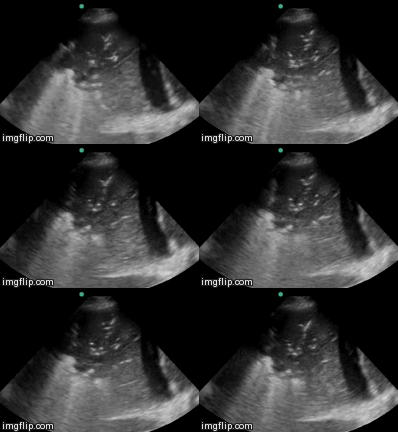
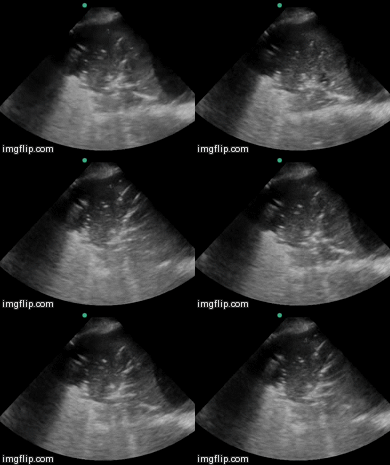
A 59 year old woman with a past medical history significant for stage IV MALT lymphoma (after chemotherapy and in remission) presented from a long term care facility for respiratory distress and altered mental status. The patient was in hypercarbic respiratory failure with a severe lactic acidosis. Her blood pressure deteriorated, she was begun on vasopressors and intubated. Pertinent labs demonstrated a white blood cell count of 0.9 X10⁶ /ml, a hemoglobin of 7.1 g/dl, and a platelet count 66 X106 /ml. The patient was started on Cefepime and Linezolid presumptively for septic shock. Ultrasounds of her thorax were performed (Videos 1 & 2).

Video 1. Ultrasound of the right thorax in the mid-axillary line.

Video 2. Ultrasound of the right thorax in the mid-axillary line (slightly more caudad).
What is the best explanation for the ultrasound findings shown above? (Click on the correct answer for an explanation)
Reference as: Aslam K, Boivin M. Ultrasound for critical care physicians: tiny bubbles. Southwest J Pulm Crit Care. 2015;10(5):216-9. doi: http://dx.doi.org/10.13175/swjpcc067-15 PDF