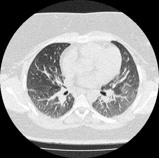
Figure 1. Thoracic CT scan in lung windows showing non-specific interstitial disease secondary to dermatomyositis.

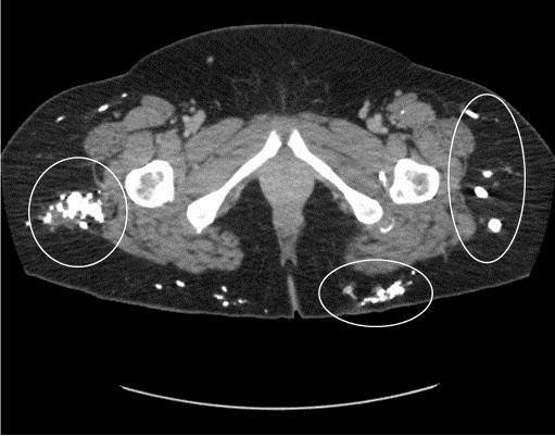
Figure 2. Pelvic CT scan showing subcutaneous calcifications (encircled).
A 36-year old woman was referred to our Interstitial Lung Disease (ILD) clinic for evaluation of dyspnea. A high-resolution CT scan of the chest showed perivascular reticular and ground glass opacities with air trapping, consistent with non-specific interstitial pneumonitis (Figure 1). She was diagnosed with connective tissue associated ILD. On review of previous images extensive subcutaneous calcifications were seen (Figure 2).
Calcinosis is an uncommon manifestation of dermatomyositis in adults (1). It is usually seen around areas of frequent trauma like the hands and elbows. In her case, a pelvic inflammatory disease may have been a trigger for this calcinosis. Calcinosis is a difficult complication to treat with some success seen with diltiazem, aluminum hydroxide, and even alendronate in children. Surgical excision may be required in some cases.
Bhupinder Natt MD
Division of Pulmonary, Allergy, Critical Care and Sleep
Banner-University Medical Center, Tucson (AZ)
Reference
- Chander S, Gordon P. Soft tissue and subcutaneous calcification in connective tissue diseases. Curr Opin Rheumatol. 2012 Mar;24(2):158-64. [CrossRef] [PubMed]
Cite as: Natt B. Medical image of the week: subcutaneous calcification in dermatomyositis. Southwest J Pulm Crit Care. 2016;13(6):317-8. doi: https://doi.org/10.13175/swjpcc130-16 PDF