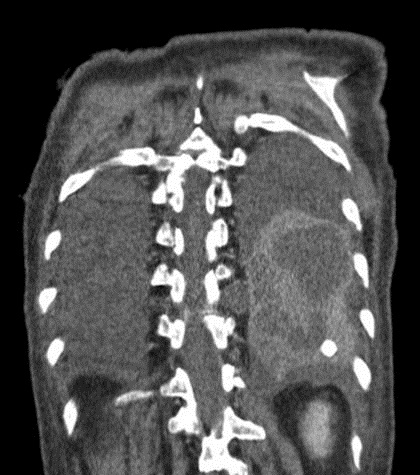
Figure 1. CT coronal view showing a left lower lobe lung abscess measuring approximately 8 x 5 cm.
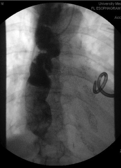
Figure 2. Barium swallow study showed dilated esophagus with tapering off at the lower esophageal sphincter junction, demonstrating the classic bird-beak like appearance.
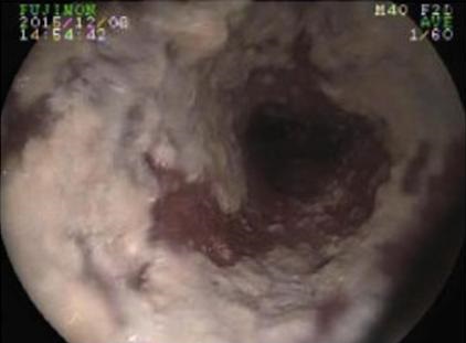
Figure 3. Upper endoscopy showing diffuse whitish plaque suggestive of candidiasis likely due to chronic stasis of food.
An 80-year old woman with past medical history of high grade serous fallopian tube carcinoma presented with 2 months history of productive cough. This was associated with shortness of breath and subjective fever, chills and weight loss of 5 pounds over 2 months. She was treated with outpatient antibiotics without improvement of symptoms. Patient was afebrile on presentation, hemodynamically stable, and saturating at 99% on room air. Lung examinations revealed dullness on percussion of left lower lung field and reduced breath sounds on the same area.
Computed tomographic imaging revealed a large lung abscess on left lower lobe (Figure 1) and moderately dilated esophagus and fluid filled to the level of gastro-esophagus junction. Barium swallow study showed a classic bird-beak like appearance (Figure 2). There was no contrast that passed through the gastro-esophagus junction during the entire course of the barium study. Upper endoscopy was performed to rule out intraluminal pathology that may contribute to the obstruction which revealed a large amount of barium and retained food in the entire esophagus with diffuse whitish plaque suggestive of candidiasis and a benign appearing intrinsic mild stenosis at lower third of esophagus (Figure 3). Pneumatic dilation and botulinum toxin injection were performed and she was started on pantoprazole. She was also started on broad-spectrum antibiotics (vancomycin, cefepime, metronidazole) for the lung abscess. A chest tube was inserted under computed tomography (CT) guidance. Subsequently, cultures from the chest tube drainage grew Streptococcus intermedius. She was discharged to a skilled nursing facility with additional 3-weeks of ampicillin-sulbactam. Repeat imaging at 3-weeks showed improvement of the lung abscess.
Achalasia is a rare primary esophageal motor disorder, with incidence of approximately 1 in 100,000 people annually and prevalence of 10 in 100,000 (1). Common presentations of achalasia includes gradual dysphagia to solid and liquids, heartburn symptoms unrelieved by adequate proton pump inhibitor therapy and weight loss. Achalasia presenting with respiratory symptoms without dysphagia is rare as this disease entity is gradual and patient will normally present with different degrees of dysphagia or regurgitation of food. This case report is a good reminder that aspiration should be considered as a cause for pneumonia in the elderly. Our patient could have been aspirating for a period of time, leading to the development of a large lung abscess. Kikuchi et al. (2) demonstrated the high incidence of silent aspiration in the elderly population. A more detailed assessment by trained swallowing therapist may aid in detecting dysphagia.
Kai Rou Tey MD1 and Naser Mahmoud MD2
1Department of Internal Medicine University of Arizona College of Medicine- South Campus
2Department of Pulmonary, Critical Care, Allergy and Sleep, University of Arizona College of Medicine
Tucson, AZ USA
References
- Francis DL, Katzka DA. Achalasia: update on the disease and its treatment. Gastroenterology. 2010 Aug;139(2):369-74. [CrossRef] [PubMed]
- Kikuchi R, Watabe N, Konno T, Mishina N, Sekizawa K, Sasaki H. High incidence of silent aspiration in elderly patients with community-acquired pneumonia. Am J Respir Crit Care Med. 1994 Jul;150(1):251-3. [CrossRef] [PubMed]
Cite as: Tey KR, Mahmoud N. Medical image of the week: achalasia with lung abscess. Southwest J Pulm Crit Care. 2016 May;12(5):194-6. doi: http://dx.doi.org/10.13175/swjpcc025-16 PDF