
Figure 1. PA (A) and lateral (B) chest X-ray showing a 5x4 cm round mass with sharp margins in retrocardiac area.
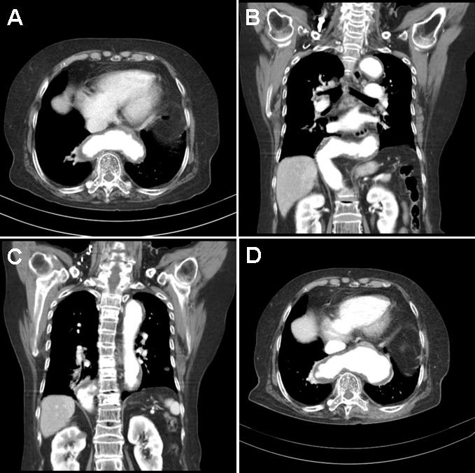
Figure 2. A-C: Initial CT image showing thoracic aorta acutely angulated above the diaphragm and crossing to the right side of the chest. Then the aorta acutely angulates again and descends into the abdomen on the right. D: Follow-up CT image after 2 years showing saccular dilatation of transverse area of thoracic aorta.
An 83-year-old female presented with epigastric discomfort and nausea for 1 month. Her past medical history included hypertension and osteoarthritis. Her vital signs at were unremarkable. Her electrocardiogram revealed only atrial premature beats. Laboratory examination, including complete blood count, liver function test, blood urea nitrogen, creatinine, and electrolytes were normal.
Esophagogastroduodenoscopy revealed minimal changes of reflux esophagitis, erosive gastritis, and extrinsic compression of lower esophagus. Her chest x-ray (Figure 1) showed a 5x4 cm sized round retrocardiac mass with sharp margin. Chest CT was ordered to evaluate the lung mass and it revealed that acutely angulated lower thoracic aorta which crossed from left to right above the left diaphragm (Figure 2). After treatment with a proton pump inhibitor and a gastrointestinal pro-motility agent, her symptoms gradually decreased. Follow-up CT after 2 years shows saccular dilatation of the transverse area of thoracic aorta (Figure 2D), however, she has no specific symptoms.
Abnormal vascular structures like a severe tortuous thoracic aorta occasionally can be confused with a lung mass or neoplasm. The most common cause of aortic disease mimicking lung mass on CXR is an aortic aneurysm (1). Some cases have reported an intervention or even an operation being performed. The symptoms of tortuosity of thoracic aorta are varied from asymptomatic to dysphagia, gastroesophageal reflux, nausea and vomiting (2). Therefore, clinical symptom is not helpful to diagnose the underlying cause. As in this case, chest computed tomography (CT) can be beneficial for the differential diagnosis between vascular lesion and lung mass. Chest CT also gives additional information for communication of the aneurysm with the aorta, relationship of vascular structure to mediastinal organs. In children, Loeys-Dietz syndrome or arterial tortuosity syndrome should be considered (3). If aortic aneurysm or tortuosity of aorta is diagnosed as a cause in older age, close observation should be performed because of the possibility of progression to aortic aneurysm, dissection or compression of adjacent organs.
Jong Seol Park, MD and Yong Sung Kim, MD, PhD
Department of Internal Medicine
Wonkwang University Sanbon Hospital
Gunpo, Korea
References
- Wixson D, Baltaxe HA, Sos TA. Pitfalls in the plain film evaluation of the thoracic aorta: the mimicry of aneurysms and adjacent masses and the value of aortography. Part I. Transverse aortic arch. Cardiovasc Radiol. 1979 Apr 27;2(2):69-76. [CrossRef] [PubMed]
- Badila E, Bartos D, Balahura C, Daraban AM. A rare cause of Dysphagia - Dysphagia aortica - complicated with intravascular disseminated coagulopathy. Maedica (Buchar). 2014 Mar;9(1):83-7. [PubMed]
- Na KJ, Park KH. Multiple aortic operations in loeys-dietz syndrome: report of 2 cases. Korean J Thorac Cardiovasc Surg. 2014 Dec;47(6):536-40. [CrossRef] [PubMed]
Cite as: Park JS, Kim YS. Medical image of the week: tortuosity of thoracic aorta mimicking a lung mass. Southwest J Pulm Crit Care. 2017;15(2):80-1. doi: https://doi.org/10.13175/swjpcc086-17 PDF