Lewis J. Wesselius, MD1
Thomas V. Colby, MD2
Departments of Pulmonary Medicine1 and Laboratory Medicine and Pathology2
Mayo Clinic Arizona
Scottsdale, AZ
History of Present Illness
A 65 year old man from Colorado presented for evaluation of “lung masses.” He had a prior diagnosis of dermatomyositis made in 2010 and had been with intravenous immunoglobulin (IVIG), prednisone and methotrexate. He had been previously seen in January, 2011 with a 5 mm left lower lobe nodule on thoracic CT which was unchanged compared to August, 2010. A thoracic CT scan done in July, 2011 in Colorado was interpreted as stable.
Over the prior month had been having chest discomfort. He had a history of pulmonary embolism (PE) and felt the pain was similar in quality to his prior PE. This prompted a chest x-ray and he was told of “lung masses”. He had also experienced 20 pound weight loss.
His current medications included methotrexate 25 mg weekly, prednisone 3 mg every other day and warfarin 7 mg daily.
PMH, SH, FH
In addition to dermatomyositis, he has a history of a left lower extremity deep venous thrombosis with PE. At that time protein S deficiency, activated protein C resistance and factor V Leiden mutation were diagnosed and an inferior vena cava filter were placed. He also has a history of paroxysmal atrial fibrillation and the prior lung nodule noted above.
He was a prior smoker, quitting in 1991, but briefly resuming in 2010. The patient had social alcohol use but no drug use.
The patient’s father died at age 75 from prostate cancer; his mother died at age 89 with heart disease; and he had a sister living with throat cancer.
Physical Examination
Vital signs: Afebrile; Blood pressure 114/65 mm/Hg; Pulse 80 regular; Oxygen Saturation 97% on room air at rest
HEENT: limited ability to open mouth
Chest: few late exp wheezes
CV: Regular rhythm, no murmur
Skin: diffuse erythema, particularly on face.
Neuro: muscle strength normal
Radiography
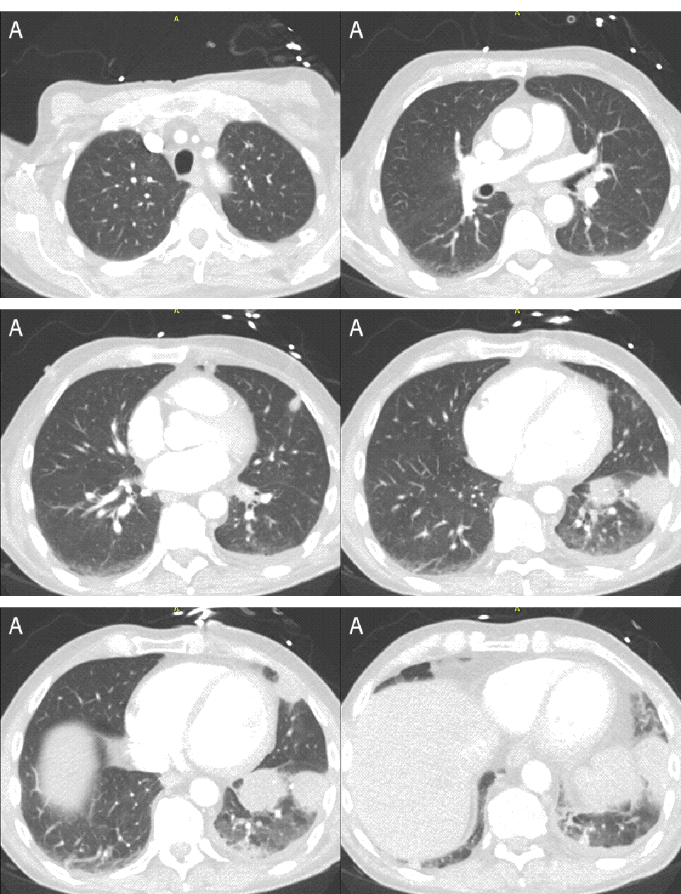
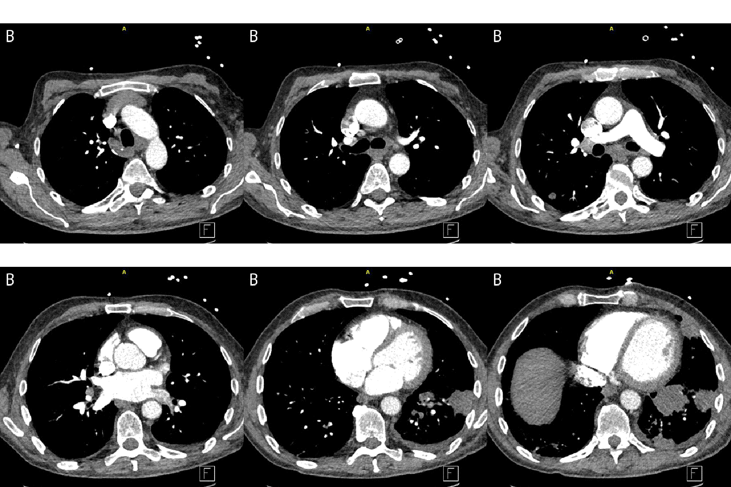
His thoracic CT is shown in Figure 1.


Figure 1. Movies of the thoracic CT scan showing lung windows (Panel A, upper panel) and mediastinal windows (Panel B, lower panel).
Which of the following are pulmonary manifestations of dermatomyositis?
- Lung cancer
- Aspiration pneumonia
- Interstitial lung disease
- Metastatic cancer particularly from the cervix, pancreas, breasts, ovaries, gastrointestinal tract and lymph nodes
- All of the above
Reference as: Wesselius LJ, Colby TV. May 2013 pulmonary case of the month: the cure can be worse than the disease. Southwest J Pulm Crit Care. 2013;6(5):199-208. PDF