Sudheer Penupolu, MD
Philip J. Lyng, MD
Lewis J. Wesselius, MD
Department of Pulmonary Medicine
Mayo Clinic Arizona
Scottsdale, AZ
History of Present Illness
A 51 year old woman was seen with a chief complaint of gradually increasing shortness of breath. She was at baseline five months prior to presentation but noticed dyspnea on minimal exertion initially at a higher altitude, gradually progressing to dyspnea at rest. She was tried on 2 courses of antibiotics with no significant improvement. In addition to the dyspnea, she has some non productive cough but no fevers.
PMH, SH, FH
She had a renal transplant in 1997 for IgA disease and has a history of type II diabetes and hypertension.
She is a life long nonsmoker and has only occasional alcohol use. She is employed as a utility designer and has no exposure to any dusts, fumes or exotic animals.
Family history is noncontributory.
Medications
- Atenolol
- Lasix
- Prednisone 2 mg q daily
- Rosuvastatin
- Sirolimus 2 mg po q daily
There have been no changes in the doses in the past few years.
Physical Examination
Physical examination reveals no abnormalities and her lung auscultation is clear.
Laboratory
Her complete blood count (CBC), urinanalysis, liver function tests, and calcium were all within normal limits.
Radiology
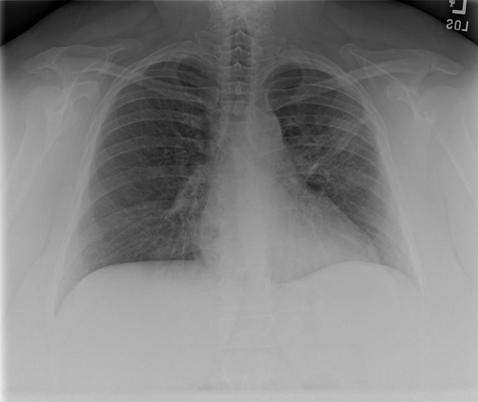
An x-ray of the chest is shown in Figure 1.

Figure 1. Initial PA chest radiograph.
Which of the below is the best interpretation of her chest x-ray?
Reference as: Penupolu S, Lyng PJ, Wesselius LJ. March 2014 pulmonary case of the month: the cure may be worse than the disease. Southwest J Pulm Crit Care. 2014;8(3):142-51. http://dx.doi.org/10.13175/swjpcc005-14 PDF