
Correct!
4. The frontal chest radiograph shows a partially circumscribed opacity projected over the right thorax
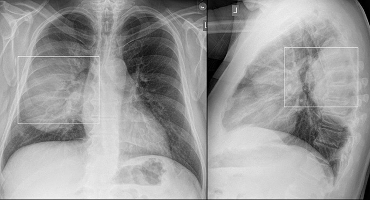
Frontal and lateral chest radiography shows a homogenous mass projected over the right upper lung. The mass shows a well-defined inferior margin and ill-defined superior margin and has extensive posterior chest wall contact. There is no evidence of cystic lung disease or pulmonary fibrosis. The mediastinal contours appear normal. An artificial intelligence (AI) computer-aided detection program applied to the chest radiograph successfully identifies and localizes the lesion (Figure 2).

Figure 2. Frontal and lateral chest radiography with the application of an artificial intelligence (Ai) computer-aided detection program shows a bounding box over the right lung mass reflecting AI detection of the abnormality. To view figure 2 in a separate, enlarged window click here.
Which of the following is correct regarding the description of the chest radiographic findings of the left-sided lesion? (click on the correct answer to be directed to the third of 11 pages)