Search Journal-type in search term and press enter
Southwest Pulmonary and Critical Care Fellowships

Critical Care

Last 50 Critical Care Postings

(Most recent listed first. Click on title to be directed to the manuscript.)

January 2025 Critical Care Case of the Month: A 35-Year-Old Admitted After
   a Fall
October 2024 Critical Care Case of the Month: Respiratory Failure in a
   Patient with Ulcerative Colitis
July 2024 Critical Care Case of the Month: Community-Acquired
   Meningitis
April 2024 Critical Care Case of the Month: A 53-year-old Man Presenting
   with Fatal Acute Intracranial Hemorrhage and Cryptogenic Disseminated
   Intravascular Coagulopathy 
Delineating Gastrointestinal Dysfunction Variants in Severe Burn Injury
   Cases: A Retrospective Case Series with Literature Review
Doggonit! A Classic Case of Severe Capnocytophaga canimorsus Sepsis
January 2024 Critical Care Case of the Month: I See Tacoma
October 2023 Critical Care Case of the Month: Multi-Drug Resistant
   K. pneumoniae
May 2023 Critical Care Case of the Month: Not a Humerus Case
Essentials of Airway Management: The Best Tools and Positioning for 
   First-Attempt Intubation Success (Review)
March 2023 Critical Care Case of the Month: A Bad Egg
The Effect of Low Dose Dexamethasone on the Reduction of Hypoxaemia
   and Fat Embolism Syndrome After Long Bone Fractures
Unintended Consequence of Jesse’s Law in Arizona Critical Care Medicine
Impact of Cytomegalovirus DNAemia Below the Lower Limit of
   Quantification: Impact of Multistate Model in Lung Transplant Recipients
October 2022 Critical Care Case of the Month: A Middle-Aged Couple “Not
   Acting Right”
Point-of-Care Ultrasound and Right Ventricular Strain: Utility in the
   Diagnosis of Pulmonary Embolism
Point of Care Ultrasound Utility in the Setting of Chest Pain: A Case of
   Takotsubo Cardiomyopathy
A Case of Brugada Phenocopy in Adrenal Insufficiency-Related Pericarditis
Effect Of Exogenous Melatonin on the Incidence of Delirium and Its 
   Association with Severity of Illness in Postoperative Surgical ICU Patients
Pediculosis As a Possible Contributor to Community-Acquired MRSA
   Bacteremia and Native Mitral Valve Endocarditis
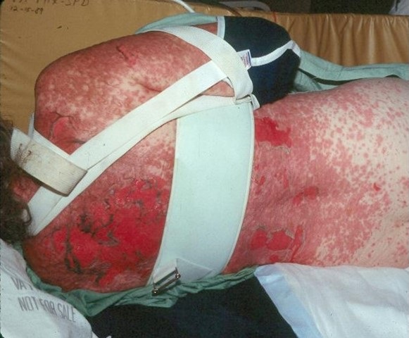
April 2022 Critical Care Case of the Month: Bullous Skin Lesions in
   the ICU
Leadership in Action: A Student-Run Designated Emphasis in
   Healthcare Leadership
MSSA Pericarditis in a Patient with Systemic Lupus
   Erythematosus Flare
January 2022 Critical Care Case of the Month: Ataque Isquémico
   Transitorio in Spanish 
Rapidly Fatal COVID-19-associated Acute Necrotizing
   Encephalopathy in a Previously Healthy 26-year-old Man 
Utility of Endobronchial Valves in a Patient with Bronchopleural Fistula in
   the Setting of COVID-19 Infection: A Case Report and Brief Review
October 2021 Critical Care Case of the Month: Unexpected Post-
   Operative Shock 
Impact of In Situ Education on Management of Cardiac Arrest after
   Cardiac Surgery
A Case and Brief Review of Bilious Ascites and Abdominal Compartment
   Syndrome from Pancreatitis-Induced Post-Roux-En-Y Gastric Remnant
   Leak
Methylene Blue Treatment of Pediatric Patients in the Cardiovascular
   Intensive Care Unit
July 2021 Critical Care Case of the Month: When a Chronic Disease
   Becomes Acute
Arizona Hospitals and Health Systems’ Statewide Collaboration Producing a
   Triage Protocol During the COVID-19 Pandemic
Ultrasound for Critical Care Physicians: Sometimes It’s Better to Be Lucky
   than Smart
High Volume Plasma Exchange in Acute Liver Failure: A Brief Review
April 2021 Critical Care Case of the Month: Abnormal Acid-Base Balance
   in a Post-Partum Woman
First-Attempt Endotracheal Intubation Success Rate Using A Telescoping
   Steel Bougie 
January 2021 Critical Care Case of the Month: A 35-Year-Old Man Found
   Down on the Street
A Case of Athabaskan Brainstem Dysgenesis Syndrome and RSV
   Respiratory Failure
October 2020 Critical Care Case of the Month: Unexplained
   Encephalopathy Following Elective Plastic Surgery
Acute Type A Aortic Dissection in a Young Weightlifter: A Case Study with
   an In-Depth Literature Review
July 2020 Critical Care Case of the Month: Not the Pearl You Were
   Looking For...
Choosing Among Unproven Therapies for the Treatment of Life-Threatening
COVID-19 Infection: A Clinician’s Opinion from the Bedside
April 2020 Critical Care Case of the Month: Another Emerging Cause
   for Infiltrative Lung Abnormalities
Further COVID-19 Infection Control and Management Recommendations for
   the ICU
COVID-19 Prevention and Control Recommendations for the ICU
Loperamide Abuse: A Case Report and Brief Review
Single-Use Telescopic Bougie: Case Series
Safety and Efficacy of Lung Recruitment Maneuvers in Pediatric Post-
   Operative Cardiac Patients
January 2020 Critical Care Case of the Month: A Code Post Lung 
   Needle Biopsy


For complete critical care listings click here.

The Southwest Journal of Pulmonary and Critical Care publishes articles directed to those who treat patients in the ICU, CCU and SICU including chest physicians, surgeons, pediatricians, pharmacists/pharmacologists, anesthesiologists, critical care nurses, and other healthcare professionals. Manuscripts may be either basic or clinical original investigations or review articles. Potential authors of review articles are encouraged to contact the editors before submission, however, unsolicited review articles will be considered.

------------------------------------------------------------------------------------

Thursday
Mar022017

March 2017 Critical Care Case of the Month

Kyle J. Henry, MD

Banner University Medical Center Phoenix

Phoenix, AZ USA


History of Present Illness

A 50-year-old man presented to the emergency room via private vehicle complaining of 5 days of intermittent chest and right upper quadrant pain. Associated with the pain he had nausea, cough, shortness of breath, lower extremity edema, and palpitations. 

Past Medical History, Social History, and Family History

He had a history of hypertension and diabetes mellitus but was on no medications and had not seen a provider in years. He was disabled from his job as a construction worker. He had smoked a pack per day for 30 years. He was a heavy daily ethanol consumer. He had an extensive family history of diabetes.

Physical Examination

  • Vitals: T 36.4 C, pulse 106/min and regular, blood pressure 96/69 mm Hg, respiratory rate 19 breaths/min, SpO2 98% on room air
  • Lungs: clear
  • Heart: regular rhythm without murmur.
  • Abdomen: mild RUQ tenderness
  • Extremities: No edema noted.

Electrocardiogram

His electrocardiogram is show in Figure 1.

Figure 1. Admission electrocardiogram.

Which of the following are true regarding the electrocardiogram? (Click on the correct answer to proceed to the second of seven pages)

  1. The lack of Q waves in V2 and V3 excludes an anteroseptal myocardial infarction
  2. The S1Q3T3 patter is diagnostic of a pulmonary embolism
  3. There are nonspecific ST and T wave changes
  4. 1 and 3
  5. All of the above

Cite as: Henry KJ. March 2017 critical care case of the month. Southwest J Pulm Crit Care. 2017;14(3):94-102. doi: https://doi.org/10.13175/swjpcc021-17 PDF

Friday
Feb032017

Ultrasound for Critical Care Physicians: Unchain My Heart

William Mansfield, MD

Michel Boivin, MD


Division of Pulmonary, Critical Care and Sleep Medicine

Department of Medicine,

University of New Mexico School of Medicine

Albuquerque, NM USA


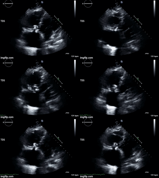
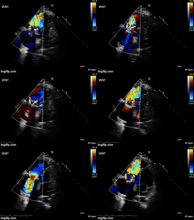
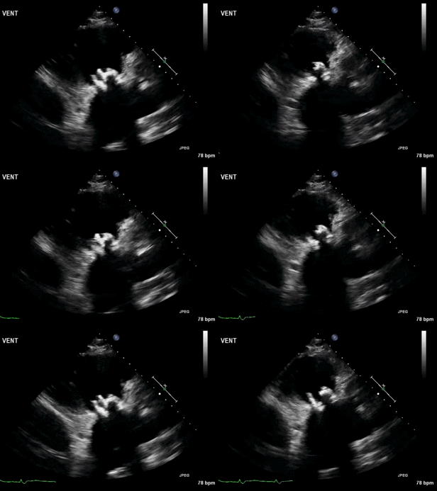
A 46-year-old man presented after a motor vehicle collision. He suffered abdominal injuries (liver laceration, avulsed gall bladder) which were successfully managed non-operatively. The patient remained intubated on mechanical ventilation and remained hypotensive after the injuries resolved. The patient required norepinephrine at low doses to maintain a normal blood pressure. It was noted the patient had a history of remote tricuspid valve replacement. A bedside echocardiogram was then performed to determine the etiology of the patient’s persistent hypotension after hypovolemia had been excluded.

Video 1. Apical four chamber view centered on the right heart.


Video 2. Apical four chamber view centered on the right heart, with color Doppler over the right atrium and ventricle.


Video 3. Right ventricular inflow view.


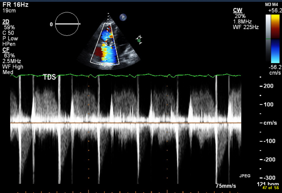
Figure 1. Continuous-wave Doppler tracing through the tricuspid valve.


What tricuspid pathology do the following videos and images demonstrate? (Click on the correct answer to proceed an explanation and discussion)

  1. Mobile vegetation
  2. Tricuspid Regurgitation
  3. Tricuspid Stenosis
  4. All of the above

Cite as: Mansfield W, Boivin M. Ultrasound for critical care physicians: unchain my heart. Southwest J Pulm Crit Care. 2017;14(2):60-4. doi: http://doi.org/10.13175/swjpcc013-17 PDF

Thursday
Feb022017

February 2017 Critical Care Case of the Month

Morgan Wong, DO

Nicholas Villalobos, MD


Department of Internal Medicine

University of New Mexico

Albuquerque, NM USA


History of Present Illness

A 68-year-old man presented to the emergency department with a one-day history of lower back pain, arthralgias, and malaise. The patient had a previous splenectomy and was concerned about influenza.

Past Medical History, Social History, and Family History

He has a history of osteoarthritis, seasonal allergies, and splenectomy. He is a nonsmoker. Family history is noncontributory.

Physical Examination

Upon admission, the patient’s vital signs were notable for a temperature of 35.3 degrees Celsius, blood pressure of 74/44 mmHg, oxygen saturation of 85% on room air with a respiratory rate of 24 breaths per minute. Physical exam was prominent for non-pitting edema of the distal upper and lower extremities, as well as diffuse macular rash of the palms and soles.

Laboratory

CBC

  • White blood cell count of 6.77 X10³ cells/uL
  • Hemoglobin of 13.8 gm/dL
  • Hematocrit of 43.7%
  • Platelet count of 19 x 103 /uL

Chemistry

  • Creatinine of 3.0 mg/dL
  • CO2 < 10 mmol/L
  • Anion gap >18 mmol/L
  • Liver function tests
  • Alanine aminotransferase (ALT) of 511 U/L
  • Aspartate aminotransferase (AST) of 529 U/L
  • Total bilirubin of 1.0 mg/dL

Coagulation

  • INR of 2.07
  • Prothromin time of 22.5 seconds
  • Partial thromoboplastin time of 82.3 seconds
  • Fibrinogen level was 71 mg/dL

Arterial blood gases

  • pH of 6.91
  • pCO2 54 mmHg
  • pO2 263
  • HCO3 of 7.7 mmol/L

Procalcitonin >200 ng/ml.

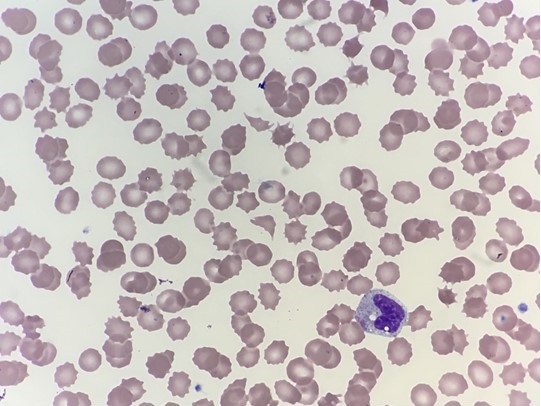
His blood peripheral smear was examined.

Figure 1: Peripheral blood smear on admission. 

Given the results of the preliminary laboratory results and peripheral smear what hematologic abnormality are you most concerned with at this time? (Click on the correct answer to proceed to the second of five pages)

  1. Autoimmune hemolytic anemia (AIHA)
  2. Disseminated intravascular coagulopathy (DIC)
  3. Microangiopathic hemolytic anemia (MAHA)
  4. Thrombotic thrombocytopenic purpura (TTP)

Cite as: Wong M, Villalobos N. February 2017 critical care case of the month. Southwest J Pulm Crit Care. 2017;14(2):54-9. doi: https://doi.org/10.13175/swjpcc144-16 PDF

Monday
Jan022017

January 2017 Critical Care Case of the Month

Seth Assar, MD

Clement U. Singarajah, MD


Pulmonary and Critical Care Medicine

Banner University Medical Center Phoenix – Phoenix

Phoenix VA Medical Center

Phoenix, AZ USA


History of Present Illness

The patient is a 48-year-old man who presented with two days of progressive shortness of breath and non-productive cough. There were no associated symptoms and the patient specifically denied fever, chills, night sweats, myalgia or other evidence of viral prodrome. He had no chest pain or tightness, nausea, vomiting, or leg swelling and he could lay flat. He had no recent travel or sick contacts and was Influenza-immunized this season.

Past Medical History

  • Hypertension
  • Hyperlipidemia
  • Type 2 diabetes mellitus with a recent hemoglobin A1C of 11%        

Social History

  • Cook at pizzeria
  • Gay and lives at home with roommate of several years
  • Smokes marijuana weekly.
  • Prior history of cocaine use

Family History

  • Noncontributory

Physical Examination

  • Vitals: T 99.1º F / HR 125 / BP 193/93 / RR 24 / SpO2 88%
  • General: Tachypneic. Alert and oriented X 4.
  • Lungs: Crackles at bases bilaterally, no wheezes
  • Heart: tachycardia
  • Abdomen: NSA
  • Skin: no needle marks or cellulitis

Laboratory

  • CBC: WBC 11,700 cells/mcL with 80% polymorphonuclear leukocytes, otherwise normal
  • Basic metabolic panel: normal
  • Brain natriuretic peptide: 120 pg/ml
  • Urine drug screen was negative for cocaine but positive for marijuana.
  • D-dimer: 0.32 mcg/mL

Hospital Course

He was admitted to the ICU but quickly deteriorated and was intubated for hypoxemia. Empiric ceftriaxone and levofloxacin were begun.

Chest x-ray demonstrated bilateral patchy airspace opacities (Figure 1).

Figure 1. Admission chest x-ray.

Which of the following should be done next? (click on the correct answer to proceed to the second of six pages)

  1. Bedside cardiac ultrasound
  2. Coccidioidomycosis serology
  3. CT scan of the chest
  4. 1 and 3
  5. All of the above

Cite as: Assar S, Singarajah CU. January 2017 critical care case of the month. Southwest J Pulm Crit Care. 2017;14(1):6-13. doi: https://doi.org/10.13175/swjpcc143-16 PDF

Friday
Dec022016

December 2016 Critical Care Case of the Month

Theodore Loftsgard APRN, ACNP

Department of Anesthesiology

Mayo Clinic Minnesota

Rochester, MN USA


Critical Care Case of the Month CME Information

Members of the Arizona, New Mexico, Colorado and California Thoracic Societies and the Mayo Clinic are able to receive 0.25 AMA PRA Category 1 Credits™ for each case they complete. Completion of an evaluation form is required to receive credit and a link is provided on the last panel of the activity. 

0.25 AMA PRA Category 1 Credit(s)™

Estimated time to complete this activity: 0.25 hours 

Lead Author(s): Theodore Loftsgard APRN, ACNP.  All Faculty, CME Planning Committee Members, and the CME Office Reviewers have disclosed that they do not have any relevant financial relationships with commercial interests that would constitute a conflict of interest concerning this CME activity.

Learning Objectives:
As a result of this activity I will be better able to:

  1. Correctly interpret and identify clinical practices supported by the highest quality available evidence.
  2. Will be better able to establsh the optimal evaluation leading to a correct diagnosis for patients with pulmonary, critical care and sleep disorders.
  3. Will improve the translation of the most current clinical information into the delivery of high quality care for patients.
  4. Will integrate new treatment options in discussing available treatment alternatives for patients with pulmonary, critical care and sleep related disorders.

Learning Format: Case-based, interactive online course, including mandatory assessment questions (number of questions varies by case). Please also read the Technical Requirements.

CME Sponsor: University of Arizona College of Medicine

Current Approval Period: January 1, 2015-December 31, 2016

Financial Support Received: None


History of Present Illness

A 62-year-old lady with primary biliary cirrhosis/autoimmune hepatitis listed for liver transplantation was admitted to the general medicine floor with progressive lethargy. She had progressive fatigue for about 10 days prior to admission. She had not been able to walk for the last few days; had anorexia; had not had a bowel movement for approximately one week; and had not taken her medicines for 4 days according to her daughter. Her family was concerned with her progressive lethargy; her darkening urine; and progressive jaundice.

She had been managed for several years on mycophenolate mofetil, budesonide, and ursodiol. She had increasing problems with ascites and had paracentesis performed about every 4 days despite taking Lasix and spironolactone. She had early encephalopathy manifested by increasing problems with word finding but had not received lactulose.

Past Medical History

She has a history of esophageal varices, recurrent cellulitis and obesity.

Physical Examination

Vital Signs: P 121 beats/min, BP 102/35 mm Hg, T 37.5 C, R 25 breaths/min

General: She was lethargic, somewhat confused but oriented to time, place and person.

Lungs: shallow respirations.

Heart: regular rhythm with a tachycardia.

Abdomen: distended with a fluid wave.

Radiography

Portable chest and abdominal x-rays were performed (Figure 1).


Figure 1. Admission chest (A) and abdominal (B) radiographs.

Which of the following best describes the x-rays? (Click on the correct answer to proceed to the second of six pages)

  1. The abdominal x-ray shows diffuse, nonspecific gaseous distention
  2. The abdominal x-ray shows gastrointestinal perforation
  3. The chest x-ray shows bilateral atelectasis
  4. The chest x-ray shows bilateral pneumonia
  5. 1 and 3
  6. 2 and 4
  7. All of the above

Cite as: Loftsgard T. December 2016 critical care case of the month. Southwest J Pulm Crit Care. 2016;13(6):278-84. doi: https://doi.org/10.13175/swjpcc104-16 PDF