Search Journal-type in search term and press enter
Southwest Pulmonary and Critical Care Fellowships
In Memoriam
Social Media

Critical Care

Last 50 Critical Care Postings

(Most recent listed first. Click on title to be directed to the manuscript.)

April 2025 Critical Care Case of the Month: Being Decisive During a 
   Difficult Treatment Dilemma 
January 2025 Critical Care Case of the Month: A 35-Year-Old Admitted After
   a Fall
October 2024 Critical Care Case of the Month: Respiratory Failure in a
   Patient with Ulcerative Colitis
July 2024 Critical Care Case of the Month: Community-Acquired
   Meningitis
April 2024 Critical Care Case of the Month: A 53-year-old Man Presenting
   with Fatal Acute Intracranial Hemorrhage and Cryptogenic Disseminated
   Intravascular Coagulopathy
Delineating Gastrointestinal Dysfunction Variants in Severe Burn Injury
   Cases: A Retrospective Case Series with Literature Review
Doggonit! A Classic Case of Severe Capnocytophaga canimorsus Sepsis
January 2024 Critical Care Case of the Month: I See Tacoma
October 2023 Critical Care Case of the Month: Multi-Drug Resistant
   K. pneumoniae
May 2023 Critical Care Case of the Month: Not a Humerus Case
Essentials of Airway Management: The Best Tools and Positioning for 
   First-Attempt Intubation Success (Review)
March 2023 Critical Care Case of the Month: A Bad Egg
The Effect of Low Dose Dexamethasone on the Reduction of Hypoxaemia
   and Fat Embolism Syndrome After Long Bone Fractures
Unintended Consequence of Jesse’s Law in Arizona Critical Care Medicine
Impact of Cytomegalovirus DNAemia Below the Lower Limit of
   Quantification: Impact of Multistate Model in Lung Transplant Recipients
October 2022 Critical Care Case of the Month: A Middle-Aged Couple “Not
   Acting Right”
Point-of-Care Ultrasound and Right Ventricular Strain: Utility in the
   Diagnosis of Pulmonary Embolism
Point of Care Ultrasound Utility in the Setting of Chest Pain: A Case of
   Takotsubo Cardiomyopathy
A Case of Brugada Phenocopy in Adrenal Insufficiency-Related Pericarditis
Effect Of Exogenous Melatonin on the Incidence of Delirium and Its 
   Association with Severity of Illness in Postoperative Surgical ICU Patients
Pediculosis As a Possible Contributor to Community-Acquired MRSA
   Bacteremia and Native Mitral Valve Endocarditis
April 2022 Critical Care Case of the Month: Bullous Skin Lesions in
   the ICU
Leadership in Action: A Student-Run Designated Emphasis in
   Healthcare Leadership
MSSA Pericarditis in a Patient with Systemic Lupus
   Erythematosus Flare
January 2022 Critical Care Case of the Month: Ataque Isquémico
   Transitorio in Spanish 
Rapidly Fatal COVID-19-associated Acute Necrotizing
   Encephalopathy in a Previously Healthy 26-year-old Man 
Utility of Endobronchial Valves in a Patient with Bronchopleural Fistula in
   the Setting of COVID-19 Infection: A Case Report and Brief Review
October 2021 Critical Care Case of the Month: Unexpected Post-
   Operative Shock 
Impact of In Situ Education on Management of Cardiac Arrest after
   Cardiac Surgery
A Case and Brief Review of Bilious Ascites and Abdominal Compartment
   Syndrome from Pancreatitis-Induced Post-Roux-En-Y Gastric Remnant
   Leak
Methylene Blue Treatment of Pediatric Patients in the Cardiovascular
   Intensive Care Unit
July 2021 Critical Care Case of the Month: When a Chronic Disease
   Becomes Acute
Arizona Hospitals and Health Systems’ Statewide Collaboration Producing a 
   Triage Protocol During the COVID-19 Pandemic
Ultrasound for Critical Care Physicians: Sometimes It’s Better to Be Lucky
   than Smart
High Volume Plasma Exchange in Acute Liver Failure: A Brief Review
April 2021 Critical Care Case of the Month: Abnormal Acid-Base Balance
   in a Post-Partum Woman
First-Attempt Endotracheal Intubation Success Rate Using A Telescoping
   Steel Bougie 
January 2021 Critical Care Case of the Month: A 35-Year-Old Man Found
   Down on the Street
A Case of Athabaskan Brainstem Dysgenesis Syndrome and RSV
   Respiratory Failure
October 2020 Critical Care Case of the Month: Unexplained
   Encephalopathy Following Elective Plastic Surgery
Acute Type A Aortic Dissection in a Young Weightlifter: A Case Study with
   an In-Depth Literature Review
July 2020 Critical Care Case of the Month: Not the Pearl You Were
   Looking For...
Choosing Among Unproven Therapies for the Treatment of Life-Threatening
   COVID-19 Infection: A Clinician’s Opinion from the Bedside
April 2020 Critical Care Case of the Month: Another Emerging Cause
   for Infiltrative Lung Abnormalities
Further COVID-19 Infection Control and Management Recommendations for
   the ICU
COVID-19 Prevention and Control Recommendations for the ICU
Loperamide Abuse: A Case Report and Brief Review
Single-Use Telescopic Bougie: Case Series
Safety and Efficacy of Lung Recruitment Maneuvers in Pediatric Post-
   Operative Cardiac Patients


For complete critical care listings click here.

The Southwest Journal of Pulmonary and Critical Care publishes articles directed to those who treat patients in the ICU, CCU and SICU including chest physicians, surgeons, pediatricians, pharmacists/pharmacologists, anesthesiologists, critical care nurses, and other healthcare professionals. Manuscripts may be either basic or clinical original investigations or review articles. Potential authors of review articles are encouraged to contact the editors before submission, however, unsolicited review articles will be considered.

------------------------------------------------------------------------------------

Thursday
Jan022014

January Critical Care Case of the Month: Bad Cough

Bhupinder Natt MD

Linda Snyder MD

Janet Campion MD


University of Arizona Medical Center

Tucson, AZ


History of Present Illness

A 41 year-old man was admitted with a five-day history of cough, shortness of breath, and fever to 102° F. He was recently diagnosed with a high-grade astrocytoma of the brain and had undergone resection followed by chemotherapy with temozomide (an alkylating agent) and radiation therapy. 

PMH

  • Renal transplantation (1993)
  • Glioblastoma (astrocytoma grade 4)
  • Crohn’s disease treated with budesonide and meselamine

Medications

  • Dexamethasone 2 mg PO BID
  • Keppra 500 mg PO BID
  • Tacrolimus 1.5 mg PO AM and 1mg PO PM
  • Mycophenolate 750 mg PO BID
  • Budesonide 3 mg PO daily
  • Meselamine 1600 mg PO TID
  • Sulfamethoxazole/trimethoprim DS PO on Mon/Wed/Fri
  • Temozolomide 75 mg IM with radiotherapy

Social History

Nonsmoker, no ethanol or recreational drugs, no recent travel, and no occupational exposures.

Physical Examination

T 38.6°C, P 112 beats/min, RR 32-40 breaths/min, BP 119/76 mm Hg, SpO2 100% on NRB

General: Fatigued, ill appearing and dyspneic.

Skin: No rash or lesions, well-healed craniotomy scar

HEENT: Dry oral mucosa, pupils and extra-ocular muscles normal

Respiratory: Reduced breath sounds, fine crackles throughout all lung fields, no wheezing

CVS: Hyperdynamic precordium, tachycardia without murmur, no elevation of jugular venous pressure (JVP), peripheral vascular exam normal.

Abdomen: Soft, non-distended, no hepato-splenomegaly, normal bowel sounds.

Lymph: No cervical lymphadenopathy

Extremities: No edema, normal muscle bulk and tone.


Laboratory

WBC 11 X 103/µL, Hemoglobin 9.8 g/dL, Hematocrit 30%, Platelets 264,000/ µL

Na+ 135 meq/L, K+ 4.2 meq/L, Cl 111 meq/L, CO2 14 mmol/L, blood urea nitrogen (BUN) 46 mg/dL, creatinine 1.7 mg/dL, glucose 132 mg/dL, calcium 10.5 mg/dL, albumin 1.5 g/dL, liver function tests-within normal limits

Prothrombin time (PT) 15 sec, international normalized ratio (INR) 1.2, partial thromboplastin time (PTT) 29.9 sec
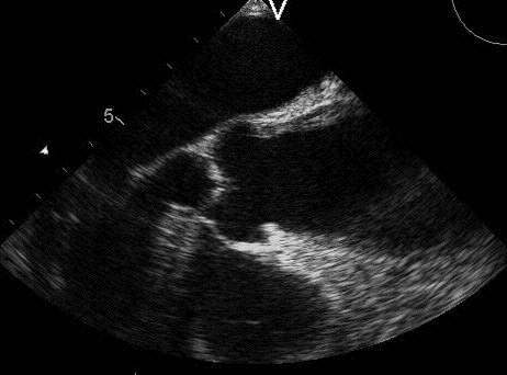
Chest X-ray

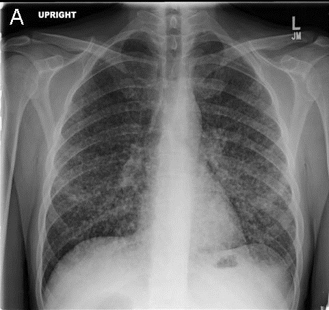
Figure 1. Admission PA (Panel A) and lateral (Panel B) chest x-ray.

What is the best description of the chest x-ray? (click on correct answer to move to next panel)

  1. Bibasilar consolidation
  2. Bilateral diffuse nodules
  3. Pneumomediastinum with subcutaneous emphysema
  4. Pulmonary edema with evidence of pulmonary hypertension
  5. Subdiaphragmatic free air

Reference as: Natt B, Snyder L, Campion J. January critical care case of the month: bad cough. Southwest J Pulm Crit Care. 2014;8(1):20-6. doi: http://dx.doi.org/10.13175/swjpcc161-13 PDF


Tuesday
Dec032013

Ultrasound for Critical Care Physicians: Unique

A 22-year-old man is seen for shortness of breath. Cardiac ultrasound / echocardiography is performed (Figure 1).

Figure 1. Cardiac ultrasound.

Which of the following best describes the ultrasound? (click on correct answer to move to next panel)

  1. Enlarged left atrium
  2. Enlarged left ventricle
  3. Enlarged right atrium
  4. Enlarged right ventricle
  5. Normal

Reference as: Gotway MB. Ultrasound for critical care physicians: unique. Southwest J Pulm Crit Care. 2013;7(6):336-7. doi: http://dx.doi.org/10.13175/swjpcc148-13 PDF

Monday
Dec022013

December 2013 Critical Care Case of the Month: I Don’t Have a Drinking Problem

Robert Raschke MD

Elijah Poulos MD

Adam Bosak MD


Critical Care Medicine

Banner Good Samaritan Medical Center

Phoenix, AZ


History of Present Illness

A 69-year-old male retired diabetic police officer was admitted to the ICU with intractable vomiting, severe abdominal pain and acute blindness. About a week prior, he suffered urinary frequency and was prescribed ciprofloxacin at urgent care with a presumptive diagnosis of urinary tract infection.  Over the course of the week his urinary frequency resolved and he became anuric, he developed progressively worsening nausea and eventually vomiting to the point that he was unable to keep anything down, and severe bilateral lower abdominal and pelvic pain.    His wife and son actually forced him into the ER when he became blind the day of admission. He denied fever, dysuria, cough and headache.   In our emergency room he was noted to be in moderate distress with tachycardia, tachypnea, hyperpnoea and completely blind in both eyes unable to discern even simple shadows.

PMH, SH, FH

The patient is a retired police officer with a past medical history of diabetes mellitus and benign prostatic hypertrophy.  The patient denied alcohol, tobacco, or illicit drug use. He works out at a local gym almost daily since being diagnosed with diabetes a couple of years ago.

Medications

  • Glipizide
  • Metformin
  • Tamsulosin

Physical Exam

Blood pressure160/95 mmHg with a heart rate of 110, respiratory rate 35, SpO2 99% on 2 lpm nasal cannula, and temp 36.0° C.  He appeared uncomfortable and moderately distressed, lethargic but arousable with GCS 13. He was able to briefly answer simple questions. His eyes were conjugate, but did not track nor fix on objects placed in front of his eyes, and he could vaguely discern the light of a bright flashlight shined into both eyes. His pupils were 3-4 mm and fixed, with no light reflex elicitable, even with magnified examination of the pupil using an ophthalmoscope.  On fundoscopic exam his discs were flat, and there were no hemorrhages or other lesions seen.  He was tachycardic but regular with normal heart tones, and a bedside echocardiogram showed good left ventricular function.  He had Kussmaul breathing with an odor of ketones and clear lungs. The lower abdomen was distended and tender, and a Foley catheter insertion returned 2 liters of yellow urine which resolved his abdominal pains.  He had no peripheral edema and his hands were cool.  The rest of his physical examination was unremarkable.

Laboratory Evaluation

Initial laboratory evaluation included a white blood count 24.3 K/mm3 with 79% segmented neutrophils and no bands, hemoglobin 14.7 g/dL; sodium 138 mmol/L;  potassium 5.1 mmol/L; chloride 92 mmol/L; and CO2 4 mmol/L, yielding an anion gap of 44 when corrected.  His BUN was 116 mg/dL; creatinine of 7.7 mg/dL.  A venous blood gas showed a pH 6.77 pCO2 17 mmHg; pO2 73 mmHg; bicarbonate of 3 mmol/L. Urinalysis showed negative leukocyte esterase, 1-5 leukocytes per HPF, glycosuria and ketonuria.

Radiology Evaluation

Admission chest x-ray is in Figure 1. 

Figure 1. Admitting chest radiograph.

Computerized tomography of the abdomen showed no urinary tract obstruction (was performed after the Foley catheter was placed) and no other significant findings. Piperacillin/tazobactam and gentamicin were started for possible urinary tract infection with sepsis.

Which of the following is the best fits the clinical presentation explaining both his metabolic abnormalities and blindness? (click on correct answer to move to next panel)

  1. Acute renal failure
  2. Alcoholic ketoacidosis
  3. Diabetic ketoacidosis
  4. Ethylene glycol ingestion
  5. Methanol ingestion

Reference as: Raschke RA, Poulos E, Bosak A. December 2013 critical care case of the month: I don't have a drinking problem. Southwest J Pulm Crit Care. 2013;7(6):328-35. doi: http://dx.doi.org/10.13175/swjpcc141-13 PDF

Sunday
Nov032013

Ultrasound for Critical Care Physicians: Atypical Headache

A 76 year old woman presented with complaints of a new onset of headache especially on the right. Brain CT scan and MRI were unremarkable. Her erythrocyte sedimentation rate was markedly elevated. An ultrasound of the chest was performed (Figures 1 and 2).

Figure 1. Long axis view through the aortic valve area (movie has been slowed).


Figure 2. Transverse view through the aorta (movie has been slowed).


What does the ultrasound show?

  1. Aortic aneurysm
  2. Aortic insufficiency
  3. Aortic stenosis
  4. Aortic wall thickening
  5. 1 and 2
  6. 1 and 4
  7. 2 and 4

Reference as: Gotway MB. Ultrasound for critical care physicians: atypical headache. Southwest J Pulm Crit Care. 2013;7(5):289-90. doi: http://dx.doi.org/10.13175/swjpcc125-13 PDF

Saturday
Nov022013

November 2013 Critical Care Case of the Month: A Series of Unfortunate Infections

Kenneth K. Sakata, MD

Karen L. Sapienza, MD

Lewis J. Wesselius, MD 


Department of Pulmonary Medicine

Mayo Clinic Arizona

Scottsdale, AZ


History of Present Illness

A 22 year old man was admitted with fever for 2 weeks. He had a history of acute lymphocytic leukemia (ALL) and had received a stem cell transplant in (SCT) in May 2013.

PMH, SH, FH

Other than the ALL and STC transplant there was no significant PMH, SH, or FH. The STC was uneventful.

Physical Examination

T 38.6°C with a pulse of 110 beats/min. Otherwise the physical examination was unremarkable.

CT scan

A CT scan of the thorax was performed in a search for the source of the fever (Figure 1).

Figure 1. Admission thoracic CT scan showing an abnormality. The remainder of the CT scan was unremarkable.

Which of the following best describes the CT abnormality?

  1. Chest wall abnormality in the left chest
  2. Focal area of consolidation in the left lung
  3. Focal area of consolidation in the right lung
  4. Mass in the left lung
  5. Mass in the right lung

Reference as: Sakata KK, Sapienza KL, Wesselius LJ. November 2013 pulmonary case of the month: a series of unfortunate infections. Southwest J Pulm Crit Care. 2013;7(5):280-8. doi: http://dx.doi.org/10.13175/swjpcc139-13 PDF