Search Journal-type in search term and press enter
Southwest Pulmonary and Critical Care Fellowships

Critical Care

Last 50 Critical Care Postings

(Most recent listed first. Click on title to be directed to the manuscript.)

October 2024 Critical Care Case of the Month: Respiratory Failure in a
   Patient with Ulcerative Colitis
July 2024 Critical Care Case of the Month: Community-Acquired
   Meningitis
April 2024 Critical Care Case of the Month: A 53-year-old Man Presenting
   with Fatal Acute Intracranial Hemorrhage and Cryptogenic Disseminated
   Intravascular Coagulopathy
Delineating Gastrointestinal Dysfunction Variants in Severe Burn Injury
   Cases: A Retrospective Case Series with Literature Review
Doggonit! A Classic Case of Severe Capnocytophaga canimorsus Sepsis
January 2024 Critical Care Case of the Month: I See Tacoma
October 2023 Critical Care Case of the Month: Multi-Drug Resistant
   K. pneumoniae
May 2023 Critical Care Case of the Month: Not a Humerus Case
Essentials of Airway Management: The Best Tools and Positioning for 
   First-Attempt Intubation Success (Review)
March 2023 Critical Care Case of the Month: A Bad Egg
The Effect of Low Dose Dexamethasone on the Reduction of Hypoxaemia
   and Fat Embolism Syndrome After Long Bone Fractures
Unintended Consequence of Jesse’s Law in Arizona Critical Care Medicine
Impact of Cytomegalovirus DNAemia Below the Lower Limit of
   Quantification: Impact of Multistate Model in Lung Transplant Recipients
October 2022 Critical Care Case of the Month: A Middle-Aged Couple “Not
   Acting Right”
Point-of-Care Ultrasound and Right Ventricular Strain: Utility in the
   Diagnosis of Pulmonary Embolism
Point of Care Ultrasound Utility in the Setting of Chest Pain: A Case of
   Takotsubo Cardiomyopathy
A Case of Brugada Phenocopy in Adrenal Insufficiency-Related Pericarditis
Effect Of Exogenous Melatonin on the Incidence of Delirium and Its 
   Association with Severity of Illness in Postoperative Surgical ICU Patients
Pediculosis As a Possible Contributor to Community-Acquired MRSA
   Bacteremia and Native Mitral Valve Endocarditis
April 2022 Critical Care Case of the Month: Bullous Skin Lesions in
   the ICU
Leadership in Action: A Student-Run Designated Emphasis in
   Healthcare Leadership
MSSA Pericarditis in a Patient with Systemic Lupus
   Erythematosus Flare
January 2022 Critical Care Case of the Month: Ataque Isquémico
   Transitorio in Spanish 
Rapidly Fatal COVID-19-associated Acute Necrotizing
   Encephalopathy in a Previously Healthy 26-year-old Man 
Utility of Endobronchial Valves in a Patient with Bronchopleural Fistula in
   the Setting of COVID-19 Infection: A Case Report and Brief Review
October 2021 Critical Care Case of the Month: Unexpected Post-
   Operative Shock 
Impact of In Situ Education on Management of Cardiac Arrest after
   Cardiac Surgery
A Case and Brief Review of Bilious Ascites and Abdominal Compartment
   Syndrome from Pancreatitis-Induced Post-Roux-En-Y Gastric Remnant
   Leak
Methylene Blue Treatment of Pediatric Patients in the Cardiovascular
   Intensive Care Unit
July 2021 Critical Care Case of the Month: When a Chronic Disease
   Becomes Acute
Arizona Hospitals and Health Systems’ Statewide Collaboration Producing a 
   Triage Protocol During the COVID-19 Pandemic
Ultrasound for Critical Care Physicians: Sometimes It’s Better to Be Lucky
   than Smart
High Volume Plasma Exchange in Acute Liver Failure: A Brief Review
April 2021 Critical Care Case of the Month: Abnormal Acid-Base Balance
   in a Post-Partum Woman
First-Attempt Endotracheal Intubation Success Rate Using A Telescoping
   Steel Bougie 
January 2021 Critical Care Case of the Month: A 35-Year-Old Man Found
   Down on the Street
A Case of Athabaskan Brainstem Dysgenesis Syndrome and RSV
   Respiratory Failure
October 2020 Critical Care Case of the Month: Unexplained
   Encephalopathy Following Elective Plastic Surgery
Acute Type A Aortic Dissection in a Young Weightlifter: A Case Study with
   an In-Depth Literature Review
July 2020 Critical Care Case of the Month: Not the Pearl You Were
   Looking For...
Choosing Among Unproven Therapies for the Treatment of Life-Threatening
   COVID-19 Infection: A Clinician’s Opinion from the Bedside
April 2020 Critical Care Case of the Month: Another Emerging Cause
   for Infiltrative Lung Abnormalities
Further COVID-19 Infection Control and Management Recommendations for
   the ICU
COVID-19 Prevention and Control Recommendations for the ICU
Loperamide Abuse: A Case Report and Brief Review
Single-Use Telescopic Bougie: Case Series
Safety and Efficacy of Lung Recruitment Maneuvers in Pediatric Post-
   Operative Cardiac Patients
January 2020 Critical Care Case of the Month: A Code Post Lung 
   Needle Biopsy
October 2019 Critical Care Case of the Month: Running Naked in the
   Park


For complete critical care listings click here.

The Southwest Journal of Pulmonary and Critical Care publishes articles directed to those who treat patients in the ICU, CCU and SICU including chest physicians, surgeons, pediatricians, pharmacists/pharmacologists, anesthesiologists, critical care nurses, and other healthcare professionals. Manuscripts may be either basic or clinical original investigations or review articles. Potential authors of review articles are encouraged to contact the editors before submission, however, unsolicited review articles will be considered.

------------------------------------------------------------------------------------

Thursday
Nov082012

Fatal Dynamic Hyperinflation Secondary to a Blood Clot Acting As a One-Way Valve at the Internal Orifice of a Tracheostomy Tube

Robert A Raschke MD MS

robert.raschke@bannerhealth.com 


Professor of Clinical Medicine

Banner Good Samaritan Regional Medical Center

Phoenix, Arizona


Abstract

We report the case of a patient who suffered fatal cardiopulmonary effects of a mobile blood clot adherent to the internal orifice of her tracheostomy tube. We believe the clot acted as a one-way valve, leading to dynamic hyperinflation and elevated intrinsic positive end expiratory pressure (iPEEP). This complication of a tracheostomy tube was suggested by clinical findings of expiratory wheezing, hypotension, increasing peak inspiratory pressure, and unusual but distinctive radiographic findings. Confirmation of one-way tracheostomy tube obstruction was difficult, even with a bronchoscopic examination. When this diagnosis is suspected, tracheostomy tube exchange should be rapidly performed.

Case Report 

The patient was a 59-year old woman who had undergone elective colostomy for symptomatic colonic atony. The patient developed a post-operative anastomotic leak, and septic shock. Despite surgical intervention and broad-spectrum antibiotics, acute respiratory distress syndrome ensued, necessitating prolonged mechanical ventilation. On the 29th day of admission, an 8.0 DCT Shiley tracheostomy tube was placed in an open procedure.

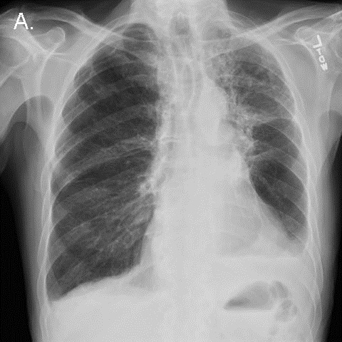
On day 33, a chest radiograph demonstrated persistent diffuse pulmonary infiltrates that had not significantly improved over the preceding 3 weeks (Figure 1).

Figure 1.  Portable chest x-ray the morning before the code arrest.

Minor bleeding was noted from the tracheostomy tube. Shortly thereafter, peak inspiratory pressures suddenly rose to the point that adequate tidal volumes could not be delivered by a mechanical ventilator. The inner cannula of the tracheostomy tube was removed. A suction catheter passed easily though the external cannula lumen, and a small amount of blood was suctioned out. However, attempts to bag-ventilate the patient became progressively more difficult. The patient's head and neck became cyanotic and mottled, and a pulse could no longer be detected. Advanced cardiac life support was initiated. Examination was significant for pan-expiratory wheezes throughout the thorax interrupted only by strenuous attempts to at bag-mask inspiration. The trachea was midline, and there was no subcutaneous crepitus. The abdomen was soft.  A bronchoscope passed through the tracheostomy tube easily, revealing a widely patent trachea and major airways. Bag ventilation transiently improved, cyanosis resolved, and a blood pressure of 150/85 was briefly obtained.  Inhaled albuterol and intravenous corticosteroids were administered.  A chest x-ray was performed (Figure 2). 

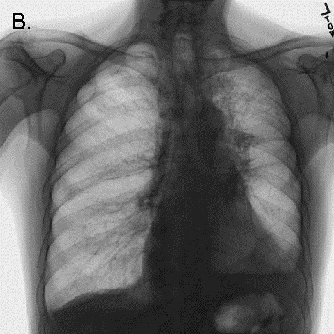
Figure 2. CXR performed during the code arrest, showing flattening of the diaphragms, and acute narrowing of the cardiac silhouette/vascular pedicle, and acute clearing of pulmonary infiltrates, consistent with hyperinflation.

Bag-ventilation became progressively more difficult, and the patient once more became hypotensive and cyanotic. The bronchoscope again passed easily through the tracheostomy and revealed the same findings as before. Needle thoracostomy was considered to treat possible pneumothorax, but the chest x-ray returned to the bedside demonstrated no evidence of barotrauma. The radiograph demonstrated striking improvement in pulmonary edema, a reduction in the size of the cardiac silhouette and vascular pedicle, and flattening of the diaphragms (see Figure 2 - note: the large radio-opacity overlying the mid-portion of the left lung is the shadow of an adherent transcutaneous pacing pad, not a pneumothorax). Further resuscitative efforts were unsuccessful.

The possibility of tracheostomy dysfunction was re-considered at some length in a postmortem debriefing. We concluded that the most likely explanation for the patient's clinical and radiological findings was dynamic hyperventilation and hemodynamic consequences of severe iPEEP induced by a dysfunction of the tracheostomy tube. 

Autopsy Findings

The tracheostomy tube was left in place, and the pathologist carefully dissected the trachea open from the carina in a caudal direction to expose the internal tip of the tracheostomy tube in-situ. A blood clot was found that nearly completely occluded the internal orifice of the tube (Figure 3, Panel A). The clot swung out of the way of some IV tubing passed inward through the external orifice of the tracheostomy tube, but swung shut again when the IV tube was removed, like a trap door (Figure 3, Panel B).

Figure 3.  Longitudinal view of the open tracheal lumen at autopsy.  Orientation: the left side of the figure is rostral. In panel A, the distal orifice of the tracheostomy tube can be seen to be nearly completely obstructed by a thrombus (black arrow). In panel B, the thrombus (black arrow) can be seen to be pushed aside by the passage of a plastic catheter (white arrow),

This clot appeared to function as a one-way valve, allowing inward passage of air, suction catheters, and a bronchoscope, but severely obstructing exhalation. We reasoned that such an obstruction could lead to wheezing and dynamic hyperinflation, and could explain the clinical and radiographic findings. Ultimately, severe iPEEP compromised cardiac preload, leading to pulselessness and death. 

No other cause for the patient's clinical syndrome was found - specifically, the patient had no antecedent history of asthma, had received no new medications on the day of the arrest, nor had any dermatological findings suggestive of anaphylaxis. The autopsy failed to reveal pulmonary embolism, mucous plugging, pneumothorax, or any histological evidence of asthma. 

Discussion

We are not the first to report dynamic hyperinflation as a complication of uni-directional tracheostomy tube obstruction (1). Several experienced clinicians at our institution recall dealing with this entity before, therefore, we suspect that it is not as rare as the paucity of clinical reports suggests. We felt that the clinical, radiological and postmortem findings in our case are sufficiently interesting, and the danger of missing this diagnosis sufficiently great, to warrant a brief review. 

Other types of tracheostomy tube dysfunction can cause high airway pressure and hypotension. Bi-directional tube obstruction from blood, dried secretions, or balloon hyperinflation is the most common (2,3). Barotrauma related to tracheostomy tubes may occur when they become displaced into the soft tissues of the neck, or into the pleural space, or when the cutaneous tracheostomy wound is sutured in an overly constrictive manner (4). 

We learned three important lessons from this unfortunate case:

  1. Clinical and radiographic findings can suggest the diagnosis of expiratory tracheostomy obstruction in a patient ventilated through a tracheostomy tube. The key clinical findings are: expiratory wheezing, hypotension, increasing iPEEP, and increasing peak inspiratory pressure.  Unexpected radiographic improvement in pulmonary edema may suggest the presence of occult iPEEP if it is not directly measured. 
  2. The diagnosis of unidirectional obstruction of a tracheostomy tube can be difficult to confirm.  The easy passage of suction catheters, or a bronchoscope, does not rule it out.  If bronchoscopy is performed emergently, the internal lumen and internal orifice of the tracheostomy tube should be examined with extreme deliberation.  This can be difficult during cardiopulmonary resuscitation.  If visualized, the potential detriment of small mobile clots should not be under-estimated.
  3. Alternative airway access should be immediately pursued in patients with tracheostomy tubes who are difficult to ventilate.  In dire clinical situations, the best diagnostic test might be to simply see if the patient improves with a new airway.  If the tracheostomy tract is likely to be mature (> 5 days old), the tracheostomy tube can simply be exchanged.  If the tract is immature, or if tube displacement is suspected, oral laryngoscopic intubation should be performed immediately.  The tracheostomy tube may need to be pulled out in order to accommodate the endotracheal tube in the trachea.  Either of these actions would likely have saved our patient's life.

References

  1. Timmus HH.  Tracheostomy: An Overview of implications, management, and morbidity.  Advances in Surgery 1973;7:199-233.
  2. Saini S, Taxak S, Singh MR.  Tracheostomy tube obstruction caused by an overinflated cuff.  Otolaryngol Head Neck Surg 2000;122:768-9.
  3. Rowe BH, Rampton J, Bota GW.  Life-threatening luminal obstruction due to mucous plugging in chronic tracheostomies: three case reports and a review of the literature.  J Emerg Med 1996;14:565-7.
  4. Tayal VS. Tracheostomies. Emerg Med Clin North Am 1994;12:707-27.

The author reports no financial support and no conflict of interest for this publication.

Reference as: Raschke RA. Fatal dynamic hyperinflation secondary to a blood clot acting as a one-way valve at the internal orifice of a tracheostomy tube. Southwest J Pulm Crit Care 2012;5:256-61. PDF

Friday
Nov022012

November 2012 Critical Care Case of the Month: I Just Can’t Do It Captain! I Can’t Get the Sats Up!

Bridgett Ronan, MD

Department of Pulmonary Medicine

Mayo Clinic Arizona

Scottsdale, AZ

History of Present Illness

A 61 year old man was seen in consultation after undergoing a laparoscopic repeat Nissen fundoplication with mesh reinforcement.  He developed worsening hypoxia postoperatively. He was initially extubated without difficulty to nasal cannula. However, he had progressive hypoxemia requiring a nonrebreathing mask, followed by BiPAP and eventually reintubation. Discussion with the surgeons revealed he had gastric contents present on intraoperative esophagogastroduodenoscopy (EGD). There was a small perforation of the fundus, with possible contamination of the peritoneum.

PMH, FH, SH

He has a long history of a paraesophageal hernia and reflux esophagitis and had previously undergone a Nissen fundoplication. There was also a history of atrial flutter and a 4.8 cm thoracic aortic aneurysm. A pre-operative echocardiogram was othewise normal. There was no remarkable family history. He was a non-drinker and non-smoker.

Physical Examination

Vital signs: Heart rate 79 beats/min, BP 95/67 mm Hg, Temperature 99.4°F, SpO2 78% on 100% FiO2.

His lungs were clear interiorly.

No murmurs or gallops were heard on cardiac auscultation.

His abdomen was post-surgical and distended but soft and nontender.

Which of the following is true regarding hypoxemia?

  1. Most hypoxia is secondary to alveolar-capillary block
  2. A normal pCO2 excludes hypoventilation as a cause of hypoxemia
  3. Low inspired FiO2 is a common cause of hypoxia in the ICU because of attaching air to the oxygen line on the ventilator.
  4. A normal chest x-ray excludes ventilation-perfusion mismatch as a cause of hypoxemia
  5. The patient’s age of 61 excludes a congenital heart lesion

Reference as: Ronan B. November 2012 critical care case of the month: I just can’t do it captain! I can’t get the sats up! Southwest J Pulm Crit Care 2012;5:235-41. PDF

Tuesday
Oct022012

October 2012 Critical Care Case of the Month

Henry Luedy, MD

Clement U. Singarajah, MD

Phoenix VA Medical Center

Phoenix, AZ

History of Present Illness

An 85 year old patient was admitted with hypotension and respiratory failure. He was intubated shortly after arrival and mechanical ventilation was begun. Fluids and vasopressors were begun for his hypotension.

PMH, SH, FH

His past medical history included peripheral vascular disease, abdominal aortic aneurysm repair, type 2 diabetes mellitus, hypertension, alcohol use, coronary artery disease, chronic obstructive pulmonary disease and hyperlipidemia.

Physical Examination

His vital signs were a temperature of 98.6 degrees F, heart rate 110 beats/min, respiratory rate 14 breaths per minute while intubated and receiving mechanical ventilation, and BP of 95/65 mmHg on vasopressors.

He was sedated. Lungs were clear and the heart had a regular rhythm without murmur or gallop. Abdominal examination was unremarkable and neurologic exam was limited because of sedation but without localizing signs. Plantar reflexes were down-going.

Admission Laboratory

Significant initial laboratory findings included a white blood cell count of 21,000 cells/μL, blood lactate level of 10 mmol/L and creatinine of 12 mg/dL. Urinanalysis showed pyuria and was positive for nitrates. At this time which of the following are diagnostic possibilities?

  1. Sepsis secondary to urinary tract infection (urosepsis)
  2. Community-acquired pneumonia
  3. Cardiogenic shock secondary to myocardial infarction
  4. Critical illness related corticosteroid insufficiency
  5. All of the above

Reference as: Luedy H, Singarajah CU. October 2012 critical care case of the month. Southwest J Pulm Crit Care 2012;5:179-85. PDF

Tuesday
Sep042012

September 2012 Critical Care Case of the Month

Robert A. Raschke, MD

Banner Good Samaritan Regional Medical Center

Phoenix, AZ

History of Present Illness

A 45 year old man was transferred from another medical center. He was found unresponsive, with muscle spasticity. After arrival at the outside medical center his vital signs were temperature 106.4 degrees F, heart rate 160 beats/min, respiratory rate 44 breaths per minute, and BP of 70/45 mm Hg. He was orally intubated for respiratory distress with induced by vecuronium.  His white blood cell count was 21,000 cells/μL. Chest x-ray showed bilateral consolidations and he was given fluids and gatifloxacin. His blood pressure improved to 130/94 and he was transferred.

PMH, SH, FH

He has a past medical history of quadriplegia at the C6 level with a history of severe back pain because syringomyelia.  He has a history of autonomic dysreflexia. Despite his disability he is quite functional working as a personal injury lawyer. He had been managed with a variety of medications including benzodiazepams, narcotics and baclofen. The later two were administered via an intrathecal pump which had been weaned over several weeks, and totally discontinued the day prior to admission. There is no history of smoking or alcohol abuse.

Physical Examination

His vital signs were temperature of 102.6 degrees F, heart rate 160 beats/min,  respiratory rate 14 breaths per minute, and BP of 130/50 mmHg.

He was paralyzed and mechanically ventilated. There was tenting of the skin and mottling of neck and knees. He had calloused hands and excoriated forearms. Lungs had diffuse rales and the heart rate was regular but rapid. A subcutaneous pump device was palpable in the left lower abdominal quadrant. There was a pressure sore on the coccyx.

Admission Laboratory and X-ray

His admission chest x-ray showed a diffuse 5-lobe consolidation. White blood cell count was elevated at 21,000 cells/μL.

At this time which of the following are diagnostic possibilities?

  1. Sepsis secondary to Staphylococcus aureus
  2. Pneumonia secondary to aspiration
  3. Neuroleptic malignant syndrome
  4. Benzodiazepam withdrawal
  5. All of the above

Reference as: Raschke RA. September 2012 critical care case of the month. Southwest J Pulm Crit Care 2012;5:121-5. (Click here for a PDF version)

Thursday
May102012

Correlation of Compliance with Central Line Associated Blood Stream Infection Guidelines and Outcomes: A Review of the Evidence

Jessica Hurley, MD1

Roxanne Garciaorr, MD1

Henry Luedy, MD1

Christan Jivcu, MD1

Emad Wissa, MD1

Joshua Jewell, MD1

Tonya Whiting, MD1

Richard Gerkin, MD1

Clement U. Singarajah, MD2

Richard A. Robbins, MD2

1Banner Good Samaritan Medical Center and 2Phoenix Pulmonary and Critical Care Medicine Research and Education Foundation, Phoenix, AZ

Abstract

Background 

Clinical practice guidelines are developed to assist in patient care but the evidence basis for many guidelines has been called into question.

Methods 

We conducted a literature review using PubMed and analyzed the overall quality of evidence and made strength of recommendation behind 8 Institute of Health Care (IHI) guidelines for prevention of central line associated blood stream infection (CLABSI). Quality of evidence was assessed by the American Thoracic Society (ATS) levels of evidence (levels I through III). We also examined data from our intensive care units (ICUs) for evidence of a correlation between guideline compliance and the development of VAP.

Results 

None of the guidelines was graded at level I. Two of the guidelines were graded at level II and the remaining 6 at level III. Despite the lack of evidence, 2 of the guidelines (hand hygiene, sterile gloves) were given a strong recommendation. Chlorhexidine and use of nonfemoral sites were given a moderate recommendation. In our ICUs compliance with the use of chlorhexidine correlated with a reduction in CLABSI (p<0.02) but the remainder did not.

Conclusions 

The IHI CLABSI guidelines are based on level II or III evidence. Data from our ICUs supported the use of chlorhexidine in reducing CLABSI. Until more data from well-designed controlled clinical trials become available, physicians should remain cautious when using current IHI guidelines to direct patient care decisions or as an assessment of the quality of care.

Introduction

The past three decades have seen the growth of numerous medical regulatory organizations. Many of these organizations have developed medical regulatory guidelines with over 10,000 listed under treatment/intervention in the National Guideline Clearinghouse (1). Many of these guidelines were rapidly adopted by healthcare organizations as a method to improve care. However, recent evidence suggests that many are based on opinion rather than randomized trials and most have not been shown to improve patient outcomes (2-5). We examined the IHI guidelines for prevention of CLABSI because these guidelines have been widely implemented despite what appeared to be a weak evidence basis (6). 

Methods

The study was approved by the Western Institutional Review Board.

Literature Search

In each instance PubMed was searched using central line associated blood stream infection which was cross referenced with each component of the CLABSI bundle (as modified by the Veterans Administration) using the following search terms: 1. hand hygiene; 2. cap (worn by inserter); 3. mask (worn by inserter); 4. sterile gloves (worn by inserter); 5. sterile gown (worn by inserter); 6. full body drape; 7. chlorhexidine used instead of povidone iodine (betadine); and 8. femoral sites not used. Additional studies were identified from the Related Citations in PubMed and the manuscript bibliographies. Each study was assessed for appropriateness to the guideline. Studies were required to be prospective and controlled in design. Only studies that used the incidence of CLABSI as a primary outcome measure were included in assessing the quality of evidence. 

Grading of level of evidence. The American Thoracic Society grading system was used to assess the underlying quality of evidence for the IHI CLABSI guidelines (7) (Table 1). A consensus was reached in each case. 

Table I. Levels of Evidence

Level of Evidence

     Definition


Level I (high)

Evidence from well-conducted, randomized controlled trials. 

Level II (moderate)


Evidence from well-designed, controlled trials without randomization (including cohort, patient series, and case-control studies). Level II studies also include any large case series in which systematic analysis of disease patterns was conducted, as well as reports of data on new therapies that were not collected in a randomized fashion.

Level III (low)


Evidence from case studies and expert opinion. In some instances, therapy recommendations come from antibiotic susceptibility data without clinical observations.


Strength of recommendations. Seven pulmonary and critical care fellows made strength of recommendations for each guideline. This was based not only the strength of evidence but also on clinical knowledge and judgment.

Guideline Compliance and CLABSI Incidence. We also assessed our ICUs for additional evidence of the effectiveness of the individual components of the CLABSI bundle. Data were collected monthly for a period of 50 months from January, 2007 through February, 2011. This was after the Veterans Administration requirements for CLABSI reporting and compliance was instituted. Diagnosis and compliance were assessed by a single quality assurance nurse using a standardized protocol (8). Compliance with each component of the bundle was analyzed individually and expressed as a percentage. This was correlated with the incidence of CLABSI during that month expressed in line-days. Each of the following was recorded by the quality assurance nurse: (1) line days, (2) number of CLABSI and (3) the number of audits or checklists completed during central line insertion and from those checklists (4) the number of times individual bundle practices were used including femoral location. In addition to data being kept locally, data was also entered into a centralized VA website. Entry into the website required completion of a learning session and a test correctly identifying CLABSI infections in case scenarios based on CDC definitions. The program included audits because the audit tool teaches critical bundle elements and facilitates communication about bundle adherence between team members.

Statistical analysis.  In some cases data were reanalyzed from original papers by Fisher’s exact test with a two-tailed comparison. For the data from our ICUs analyses were done using a Pearson correlation coefficient with a two-tailed test. Significance was defined as p<0.05. 

Results

Literature Review

Numbers of articles identified by PubMed search and used for grading the level of evidence and strength of recommendation are given in Table 2. Also included are the level of evidence and the strength of the recommendation.

Table 2.

*Includes maximum barrier compared to standard barrier studies.

Barrier Protection. Five of the guidelines (cap worn by inserter; mask worn by inserter; sterile gown worn by inserter; and full body drape on patient) can be grouped under maximal barrier precautions as originally described by Raad et al. (9). This single site study compared 343 randomized patients to have nontunneled central catheters inserted under maximal sterile barrier precautions or control precautions (sterile gloves and small drape only). The catheters were for non-emergency venous access for chemotherapy and/or bone marrow transplantation. All patients were followed for 3 months and there were a total of four catheter infections in the test group and 12 in the control group (p= 0.03, chi-square test). However, examination of Table 3 of the manuscript revealed that 6 of the 12 in the control and 3 of the 4 in the test group had colonization rather than septicemia. Furthermore, of the remaining 7 patients only 2 developed septicemia within 30 days (both in the control group at 7 and 10 days). The remaining patients developed septicemia long after any expected benefit from barrier precautions during insertion (35-98 days). Development of septicemia this long after insertion and would seem more likely to represent contamination during handling of the catheter. Eliminating those subjects with colonization alone and septicemia after 30 days leaves 2 of 167 in the control and none of 176 in the maximum barrier precautions group. Recalculation reveals no statistically significant reduction by (p=0.24). Consistent with this reanalysis, a recent randomized, multicenter trial comparing maximum sterile barrier precautions vs. standard precautions reported by Ishikawa et al. (10) did not demonstrate a reduction in CLABSI. CLABSI developed in 5 of 211 patients with the use of maximum sterile barrier precautions compared to 6 of 213 patients with standard precautions (gloves and drape, p=1.00). Combining Raad’s revised data with Ishikawa’s data did not demonstrate a statistically significant difference between maximum and standard barrier precautions (p=0.42).  

Two other studies were considered. One was a prospective but observational study by Lee et al. (11). Data from this study demonstrate a lower rate of infection with maximum sterile precautions (p=0.02) but was excluded because of the nonrandomization. Another study by Rijnders et al. (12) was a randomized study comparing maximum barrier precautions and standard precautions but with arterial lines. Maximum barrier precautions did not significantly lower the infection rate (p>0.1) but the study was excluded because it dealt with arterial rather than central venous lines.

It is also important to note that CLABSI has been directly linked to the organisms growing on the skin at the insertion site. Carrer et al. (21) found maximal sterile barrier precautions, when compared to standard care, decreased skin colonization at the insertion site for the first 48 hours (39% vs. 69%). However, within 48 hours skin colonization was no different than at 5 days. Furthermore there was no statistical difference in device colonization found between the groups (p=0.10) indicating barrier precautions did not change the rate of CLABSI. Kim and colleagues (22) found the maximal barrier precautions successfully decreased the number of gram-positive infections (p=0.05) but actually increased fungal infections (p=0.04) while having no effect on gram-negative organisms. Given the natural flora of the skin is nearly all gram-positive organisms, sole reduction of gram-positive infections alone in this study reiterates the minimal overall effect maximal barrier precautions has on CLABSI in relation to the skin organisms at the central line insertion site and suggests CLABSI often occurs in the setting of future contamination post-insertion (22).

Based on the data and a lack of clear cut rationale into a mechanism of why maximum barrier precautions should reduce CLABSI, a weak recommendation was given to each of the components of maximum barrier precautions (cap, mask, gown and drape).

Hand hygiene and sterile gloves. The effects of hand hygiene and/or sterile gloves on the development of CLABSI have not been validated in randomized controlled trials. Observational studies have demonstrated a significant decrease in the incidence of nosocomial infections with improvements in hand hygiene and use of sterile gloves (13). Decreased mortality associated with the implementation of hand hygiene dates back to Semmelweis in 1847 (14). As the simplest and least expensive means of reducing CLABSI, hand hygiene and the use of sterile gloves were strongly recommended.

Chlorhexidine. One single institution study compared 492 arterial line insertions combined with 176 central venous catheters (15). Patients were randomized to povidone iodine (227 patients), alcohol (227 patients) and chlorhexidine (224 patients) for use in insertion as well as every 48 hour cleansing of the insertion site. Seven, six and one of the patients in each group developed bacteremia respectively. Chlorhexidine use resulted in a statistically significant reduction in bacteremia if compared to the combined povidone iodine and alcohol groups. However, statistical significance was lost when analyzed on a 3x2 table or comparing povidone iodine or alcohol with chlorhexidine individually (p>0.05, all comparisons).

Mimoz et al. (16) reported a single center study in both arterial and central venous line insertions. Patients were randomly assigned to either a solution composed of 0.25% chlorhexidine gluconate, 0.025% benzalkonium chloride, and 4% benzyl alcohol or 10% povidone iodine. The same solution was used for skin disinfection from the time of catheter insertion to the time of removal of each catheter. The use of the chlorhexidine containing solution was more efficacious in preventing line related sepsis compared to povidone iodine in preventing Gram + but not Gram negative infections but there was no overall reduction of CLABSI with chlorhexidine (3 CLABSI out of 170) compared to povidone-iodine (4 CLABSI out of 145).

Another multicenter prospective, randomized, controlled trial reported by Humar et al. (17) compared 0.5% tincture of chlorhexidine to 10% povidone-iodine as cutaneous antisepsis for central venous catheter in intensive care units. Four cases of documented catheter-related bacteremia out of 193 patients were found in the chlorhexidine group compared to 5 of 181 the povidone-iodine group (p>0.05).

Combining the above studies resulted in no significant reduction with the use of chlorhexidine (8 CLABSI out of 577) compared to povidone-iodine (15 CLABSI out of 553, p=0.14).

The results of a more recent 3-year, multi-institutional, interrupted time-series design (October 2006 to September 2009), with historical control data in the pediatric intensive care unit produced differing results (18). A nested, 18-month, nonrandomized, factorial design was used to evaluate chlorhexidine scrub and chlorhexidine-impregnated sponge compliance rates. Neither was associated with a reduction in CLABSI. Due to the results only being reported in CLABSI rate per 1000 line days it was not possible to combine the data with the other studies.

One of the randomized studies showed decreased CLABSI with chlorhexidine (15). Two of the three studies showed decreased colonization as well as decreased rates of line related sepsis while the third showed decreased exit site infections (all other findings of that study did not quite make significance). All three studies were in the ICU setting, two in surgical, evaluating both central venous catheters as well as arterial lines. In this setting with likely minimal cost difference, equal to better ease of use, and smaller studies of both venous and arterial lines, the strength of recommendation was judged as moderate.

Insertion Site. One randomized study compared infections using the femoral and internal jugular sites (19). In this study the rate of CLABSI did not differ (3/313 vs. 5/313). Another study compared the femoral and subclavian sites. It also did not show a reduction with a nonfemoral site (2/127 vs. 6/100) (20). Combining the two studies did not show a significantly lower infection rate with a nonfemoral site (7/440 nonfemoral vs. 9/413 femoral, p= 0.45). Two other nonrandomized studies examined femoral compared to subclavian and internal jugular sites (23,24). One did not show a difference between the sites (23). The other, larger study showed a lower rate with non-femoral sites (24). Three other studies were considered but were found to be nonrandomized (25-27). However, complications appear higher with the femoral route including thrombosis and hematomas (28,29).

The one observational study and the higher rate of non-infectious complications resulted in the group recommending a non-femoral site when possible. The strength of this recommendation was judged as moderate.

Guideline Compliance CLABSI Incidence. In our ICUs, 1133 audits representing 11470 line-days were assessed monthly (Appendix 1). An average of 1.3 CLABSI infections/1000 line-days occurred. Correlation between the monthly compliance with each component of the CLABSI bundle with the monthly CLABSI incidence revealed only chlorhexidine use was associated with reduced CLABSI (r=-0.35, p=0.01).

Discussion

This manuscript questions the validity of the CLABSI bundles as proposed by the IHI. We found that a systematic review of the literature revealed predominantly weak evidence to support these guidelines. Only one guideline (chlorhexidine) was supported by a randomized trial (15). However, data from our own ICUs showed a correlation between use of chlorhexidine and a reduction in CLABSI.

The diagnosis of CLABSI is difficult, requiring clinical judgment even in the presence of objective clinical criteria (8). The difficulty in diagnosis, along with the negative consequences for failure to follow the IHI guidelines, makes before and after comparisons of the incidence of CLABSI unreliable. Therefore, we sought evidence for the effectiveness of CLABSI prevention guidelines reasoning that the better the compliance with the guidelines, the lower the incidence of CLABSI. We were unable to show that improved CLABSI guideline compliance correlated with a reduced incidence of CLABSI with the exception of use of chlorhexidine.

Particularly disappointing is the data on maximum barrier precautions and reduction in CLABSI. The evidence presented in the first randomized trial was weak (9). Furthermore, when we carefully examined the data we found inclusion of catheter colonization and delays in diagnosis of over 30 days in the time from catheter insertion. It seems unlikely that contamination at the time of insertion would take over 30 days to present with sepsis. If the insertion technique was faulty (e.g., no barrier use) the infection should present within a matter of days not weeks. Many studies conflate catheter colonization with a true catheter related infection. The two entities are managed quite differently and thus need to be carefully separated (8). Reanalysis of the data eliminating the colonized patients and those who took over 30 days to present showed no reduction in CLABSI with maximum barrier protection. A more recent randomized, multicenter study would support the conclusion that there is no significant difference between maximum and standard barrier precautions (10).

We could find no randomized studies of hand hygiene and gloves in the context of CLABSI prevention. However, studies in the operating room and the intensive care unit have both demonstrated that hand hygiene decreases infection (13). Both have become standards of practice. Therefore, our group felt ardently that this should be a strong recommendation.

Use of povidone iodine or chlorhexidine is largely dependent on what is stocked at the time of central line insertion. We are unaware of data supporting physician preference for povidone iodine over chlorhexidine; in fact, our group almost universally prefers chlorhexidine. Although the evidence basis for chlorhexidine over povidone iodine is marginal, it seems reasonable to use chlorhexidine until the time that additional data are available, and therefore, chlorhexidine use was given a moderate recommendation.

The data using non-femoral sites showed no clear cut reduction in CLABSI. The femoral site may have advantages particularly in emergency situations including ease of placement, compressibility and being distant from the head and neck during resuscitation. However, it appears to come at a higher price of both hematomas and thrombosis (19,20). Based on this our group felt a moderate recommendation was justified in nonemergent situations.

Our study has several limitations. No literature review is totally comprehensive. It is possible that studies relevant to the IHI CLABSI guidelines, especially those written in a foreign language, were not identified. Second, the Phoenix VA data may be underpowered to show a small beneficial effect despite having over 11,000 line-days. Third, as with other healthcare facilities, the CLABSI guidelines at our institution were mandated and monitored. The threat of negative consequences may have compromised the objective assessment of the self-reported data, likely invalidating a before and after comparison. Fourth, correlation between guideline compliance and CLABSI incidence is not a substitute for a randomized trial. Unfortunately, the later is not possible given that guideline compliance is mandated.

In the above context, this report both confirms some aspects but differs in others from a recent report by the Veterans Administration (30). In this report the VA reported data from all ICUs and found a reduction in CLABSI and an increase in compliance from 2006-9. Although the database is much larger than the data in this report from a single institution, it suffers from the same weaknesses as our data. Not reported are the death or morbidity rates from CLABSI. Interestingly, CLABSI rates were much lower in smaller hospitals (level 4). Whether these hospitals had increased compliance was not reported, but these smaller hospitals are known to have higher all cause mortality, surgical mortality and surgical morbidity (31).

Guidelines have taken on the aura of law which is substituted for clinical judgment. For example, a nurse practitioner attempted to prevent a senior critical care physician in one of our facilities from inserting a central line because the physician was not wearing a cap. However, since the patient had no line access and was in extremis, the physician decided to proceed. Other examples are the decisions to place a femoral catheter during emergencies, when other venous access is unavailable, when a pneumothorax might be catastrophic, or when major bleeding is a risk (the femoral vein is compressible). Clinical judgment might weigh the risks of internal jugular or subclavian insertions compared to the femoral vein and conclude that the femoral site might be the best choice for the patient.

It is unclear why the IHI guidelines have received such wide acceptance given their weak evidence basis. Agencies involved in guideline writing should show restraint in guideline formulation based on opinion or weak or conflicting evidence. Only those interventions based on strong evidence which can make a real difference to patients should be designated as guidelines.

Acknowledgements

The authors acknowledge Janice Allen, MSN, RN who collected the CLABSI data reported from the Phoenix VA.

References

  1. http://www.guideline.gov/ Accessed 12-1-2011.
  2. Lee DH, Vielemeyer O. Analysis of overall level of evidence behind infectious diseases society of America practice guidelines. Arch Intern Med 2011;171:18-22.
  3. Kett DH, Cano E, Quartin AA, Mangino JE, Zervos MJ, Peyrani P, Cely CM, For KD, Scerpella EG, Ramirez JA. Implementation of guidelines for management of possible multidrug-resistant pneumonia in intensive care: an observational, multicentre cohort study. Lancet Infect Dis 2011;11:181-9.
  4. Padrnos L, Bui T, Pattee JJ, Whitmore EJ, Iqbal M, Lee S, Singarajah CU, Robbins RA. Analysis of overall level of evidence behind the Institute of Healthcare Improvement ventilator-associated pneumonia guidelines. Southwest J Pulm Crit Care 2011;3:40-8.
  5. Robbins RA, Gerkin R, Singarajah CU. Relationship between the Veterans Healthcare Administration Hospital Performance Measures and Outcomes. Southwest J Pulm Crit Care 2011;3:92-133.
  6. http://www.ihi.org/explore/centrallineinfection/Pages/default.aspx (accessed 12-1-11).
  7. Schünemann H, Jaeschke R, Cook DJ, et al. An Official ATS Statement: Grading the Quality of Evidence and Strength of Recommendations in ATS Guidelines and Recommendations. Am J Resp Crit Care Med 2006;174:605-14.
  8. Horan TC, Andrus M, Dudeck MA. CDC/NHSN surveillance definition of health care-associated infection and criteria for specific types of infections in the acute care setting. Am J Infect Control 2008;36:309-32.
  9. Raad II, Hohn DC, Gilbreath BJ, Suleiman N, Hill LA, Bruso PA, Marts K, Mansfield PF, Bodey GP. Prevention of central venous catheter-related infections by using maximal sterile barrier precautions during insertion. Infect Control Hosp Epidemiol 1994;15:231-8.
  10. Ishikawa Y, Kiyama T, Haga Y, Ishikawa M, Takeuchi H, Kimura O, Harihara Y, Sunouchi K, Furuya T, Kimura M. Maximal sterile barrier precautions do not reduce catheter-related bloodstream infections in general surgery units: a multi-institutional randomized controlled trial. Ann Surg 2010 ;251:620-3.
  11. Lee D-h, Jung KY, Choi Y-H. Use of maximal sterile barrier precautions and/or antimicrobial- coated catheters to reduce the risk of central venous catheter–related bloodstream infection. Infect Control Hosp Epidemiol 2008;29:947-50.
  12. Rijnders BJ, Van Wijngaerden E, Wilmer A, Peetermans WE. Use of full sterile barrier precautions during insertion of arterial catheters: a randomized trial. Clin Infect Dis 2003;36:743-8.
  13. Cork DP, Maxwell PJ 4th, Yeo CJ. Remembering Semmelweis: hand hygiene and its importance on today's clinical practice. Am Surg 2011;77:123-5.
  14. Maki DG, Ringer M, Alvarado CJ. Prospective randomised trial of povidone-iodine, alcohol, and chlorhexidine for prevention of infection associated with central venous and arterial catheters. Lancet. 1991;338:339-43.
  15. Mimoz O, Pieroni L, Lawrence C, Edouard A, Costa Y, Samii K, et al. Prospective, randomized trial of two antiseptic solutions for prevention of central venous or arterial catheter colonization and infection in intensive care unit patients. Crit Care Med 1996;24:1818-23.
  16. Humar A, Ostromecki A, Direnfeld J, Marshall JC, Lazar N, Houston PC, Boiteau P, Conly JM. Prospective randomized trial of 10% povidone-iodine versus 0.5% tincture of chlorhexidine as cutaneous antisepsis for prevention of central venous catheter infection. Clin Infect Dis. 2000;31:1001-7.
  17. Parienti JJ, Thirion M, Mégarbane B, Souweine B, Ouchikhe A, Polito A, Forel JM, Marqué S, Misset B, Airapetian N, Daurel C, Mira JP, Ramakers M, du Cheyron D, Le Coutour X, Daubin C, Charbonneau P; Members of the Cathedia Study Group. Femoral vs jugular venous catheterization and risk of nosocomial events in adults requiring acute renal replacement therapy: a randomized controlled trial. JAMA 2008;299:2413-22.
  18. Merrer J, De Jonghe B, Golliot F, Lefrant JY, Raffy B, Barre E, Rigaud JP, Casciani D, Misset B, Bosquet C, Outin H, Brun-Buisson C, Nitenberg G; French Catheter Study Group in Intensive Care. Complications of femoral and subclavian venous catheterization in critically ill patients: a randomized controlled trial. JAMA 2001;286:700-7.
  19. Carrer S, Bocchi A, Bortolotti M, Braga N, Gilli G, Candini M, Tartari S. Effect of different sterile barrier precautions and central venous catheter dressing on the skin colonization around the insertion site. Minerva Anestesiol 2005;71:197-206.
  20. Kim JS. Holtom P. Vigen C. Reduction of catheter-related bloodstream infections through the use of a central venous line bundle: epidemiologic and economic consequences. American Journal of Infection Control 2011;39:640-6.
  21. Deshpande KS, Hatem C, Ulrich HL, Currie BP, Aldrich TK, Bryan-Brown CW, et al. The incidence of infectious complications of central venous catheters at the subclavian, internal jugular, and femoral sites in an intensive care unit population. Critical Care Medicine 2005;33:13-20.
  22. Lorente L, Henry C, Martin MM, Jimenez A, Mora ML. Central venous catheter-related infection in a prospective and observational study of 2,595 catheters. Critical Care (London, England) 2005;9:R631-5.
  23. Casanegra AI, Brannan S, Dadu R, Ong P, Berner M, Spinning K, Hughes T, Marrinan G, Zinn K, Manthous C. Short-term femoral vein catheterization rarely causes thrombosis or bacteremia. J Hosp Med 2011;6:33-6.
  24. Hsu JF, Tsai MH, Huang HR, Lien R, Chu SM, Huang CB. Risk factors of catheter-related bloodstream infection with percutaneously inserted central venous catheters in very low birth weight infants: a center's experience in Taiwan. Pediatr Neonatol 2010;51:336-42.
  25. Tsai MH, Lien R, Wang JW, Huang HR, Chiang CC, Chu SM, Hsu JF, Huang YC. Complication rates with central venous catheters inserted at femoral and non-femoral sites in very low birth weight infants. Pediatr Infect Dis J 2009;28:966-70.
  26. Trottier SJ, Veremakis C, O’Brien J, Auer AI. Femoral deep vein thrombosis associated with central venous catheterization: results from a prospective, randomized trial. Crit Care Med 1995;23:52-9.
  27. Ge X, Cavallazzi R, Li C, Pan SM, Wang YW, Wang FL. Central venous access sites for the prevention of venous thrombosis, stenosis and infection. Cochrane Database Syst Rev 2012;3:CD004084.
  28. Render ML, Hasselbeck R, Freyberg RW, Hofer TP, Sales AE, Almenoff PL. Reduction of central line infections in Veterans Administration intensive care units: an observational cohort using a central infrastructure to support learning and improvement. BMJ Qual Saf 2011;20:725-732.
  29. Robbins RA, Gerkin R, Singarajah CU. Relationship between the veterans healthcare administration hospital performance measures and outcomes. Southwest J Pulm Crit Care 2011;3:92-133.

Refernce as: Hurley J, Garciaorr R, Luedy H, Jivcu C, Wissa E, Jewell J, Whiting T, Gerkin R, Singarajah CU, Robbins RA. Correlation of compliance with central line associated blood stream infection guidelines and outcomes: a review of the evidence. Southwest J Pulm Crit Care 2012;4:163-73. (Click here for a PDF version of manuscript)