
July 2017 Pulmonary Case of the Month
Robert W. Viggiano, MD
Department of Pulmonary Medicine
Mayo Clinic Arizona
Scottsdale, AZ USA
History of Present Illness
The patient is a 19-year-old woman who went to a local Emergency Room 12/23/15 for chest pain she described as pleurisy. She was told she had pneumonia and a chest x-ray was reported to show a lingular infiltrate (Figure 1).
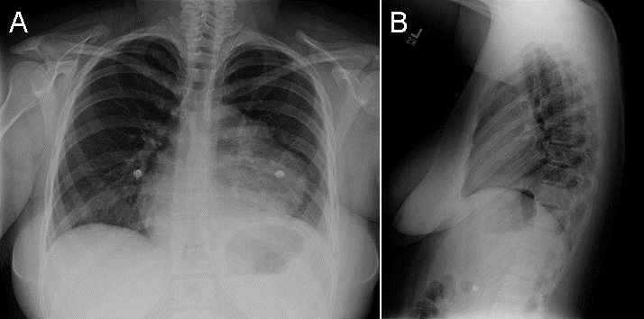
Figure 1. PA (A) and lateral (B) chest radiograph taken 12/23/15.
She was treated with antibiotics and improved. She was well until 9/2/16 when she again returned to the emergency room complaining of hemoptysis. A chest x-ray was reported as showing a lingular infiltrate (Figure 2).
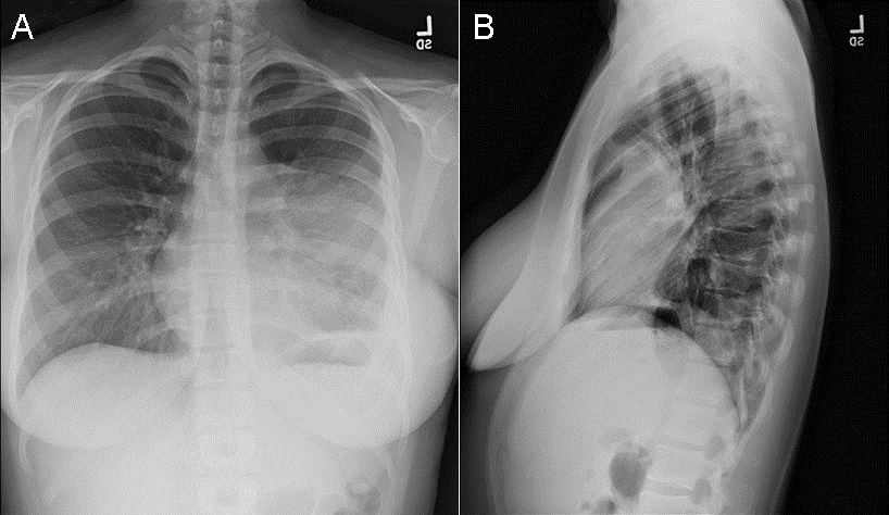
Figure 2. PA (A) and lateral (B) chest radiograph taken 9/2/16.
She was treated with azithromycin but her cough persisted sometimes with a small amount of blood in her sputum. She was referred because of her persistent symptoms and her abnormal chest x-ray.
Past Medical History, Social History and Family History
- She is now taking fluoxetine daily.
- She has a history of pediatric autoimmune neuropsychiatric disorder associated with Group A Streptococcus and was treated with antibiotics for 4-5 years.
- Nonsmoker.
Physical Examination
Her physical examination was unremarkable.
Which of the following are true? (Click on the correct answer to proceed to the second of five pages)
- Her chest radiographs are consistent with pneumonia
- Lung cancer is an unlikely consideration in a 19-year-old
- The chest x-ray findings represent a well-known complication of pediatric autoimmune neuropsychiatric disorder
- 1 and 3
- All of the above
Cite as: Viggiano RW. July 2017 pulmonary case of the month. Southwest J Pulm Crit Care. 2017;15(1):1-6. doi: https://doi.org/10.13175/swjpcc082-17 PDF
 Post a Comment
Reader Comments