Search Journal-type in search term and press enter
Southwest Pulmonary and Critical Care Fellowships

Pulmonary

Last 50 Pulmonary Postings

(Click on title to be directed to posting, most recent listed first)

Glucagon‐like Peptide-1 Agonists and Smoking Cessation: A Brief Review
September 2024 Pulmonary Case of the Month: An Ounce of Prevention
   Cased a Pound of Disease
Yield and Complications of Endobronchial Ultrasound Using the Expect
   Endobronchial Ultrasound Needle
June 2024 Pulmonary Case of the Month: A Pneumo-Colic Association
March 2024 Pulmonary Case of the Month: A Nodule of a Different Color
December 2023 Pulmonary Case of the Month: A Budding Pneumonia
September 2023 Pulmonary Case of the Month: A Bone to Pick
A Case of Progressive Bleomycin Lung Toxicity Refractory to Steroid Therapy
June 2023 Pulmonary Case of the Month: An Invisible Disease
February 2023 Pulmonary Case of the Month: SCID-ing to a Diagnosis
December 2022 Pulmonary Case of the Month: New Therapy for Mediastinal
   Disease
Kaposi Sarcoma With Bilateral Chylothorax Responsive to Octreotide
September 2022 Pulmonary Case of the Month: A Sanguinary Case
Electrotonic-Cigarette or Vaping Product Use Associated Lung Injury:
   Diagnosis of Exclusion
June 2022 Pulmonary Case of the Month: A Hard Nut to Crack
March 2022 Pulmonary Case of the Month: A Sore Back Leading to 
   Sore Lungs
Diagnostic Challenges of Acute Eosinophilic Pneumonia Post Naltrexone
   Injection Presenting During The COVID-19 Pandemic
Symptomatic Improvement in Cicatricial Pemphigoid of the Trachea 
   Achieved with Laser Ablation Bronchoscopy
Payer Coverage of Valley Fever Diagnostic Tests
A Summary of Outpatient Recommendations for COVID-19 Patients
   and Providers December 9, 2021
December 2021 Pulmonary Case of the Month: Interstitial Lung
   Disease with Red Knuckles
Alveolopleural Fistula In COVID-19 Treated with Bronchoscopic 
   Occlusion with a Swan-Ganz Catheter
Repeat Episodes of Massive Hemoptysis Due to an Anomalous Origin 
   of the Right Bronchial Artery in a Patient with a History
   of Coccidioidomycosis
September 2021 Pulmonary Case of the Month: A 45-Year-Old Woman with
   Multiple Lung Cysts
A Case Series of Electronic or Vaping Induced Lung Injury
June 2021 Pulmonary Case of the Month: More Than a Frog in the Throat
March 2021 Pulmonary Case of the Month: Transfer for ECMO Evaluation
Association between Spirometric Parameters and Depressive Symptoms 
   in New Mexico Uranium Workers
A Population-Based Feasibility Study of Occupation and Thoracic 
   Malignancies in New Mexico
Adjunctive Effects of Oral Steroids Along with Anti-Tuberculosis Drugs
   in the Management of Cervical Lymph Node Tuberculosis
Respiratory Papillomatosis with Small Cell Carcinoma: Case Report and
   Brief Review
December 2020 Pulmonary Case of the Month: Resurrection or 
   Medical Last Rites?
Results of the SWJPCC Telemedicine Questionnaire
September 2020 Pulmonary Case of the Month: An Apeeling Example
June 2020 Pulmonary Case of the Month: Twist and Shout
Case Report: The Importance of Screening for EVALI
March 2020 Pulmonary Case of the Month: Where You Look Is 
   Important
Brief Review of Coronavirus for Healthcare Professionals February 10, 2020
December 2019 Pulmonary Case of the Month: A 56-Year-Old
   Woman with Pneumonia
Severe Respiratory Disease Associated with Vaping: A Case Report
September 2019 Pulmonary Case of the Month: An HIV Patient with
   a Fever
Adherence to Prescribed Medication and Its Association with Quality of Life
Among COPD Patients Treated at a Tertiary Care Hospital in Puducherry
    – A Cross Sectional Study
June 2019 Pulmonary Case of the Month: Try, Try Again
Update and Arizona Thoracic Society Position Statement on Stem Cell 
   Therapy for Lung Disease
March 2019 Pulmonary Case of the Month: A 59-Year-Old Woman
   with Fatigue
Co-Infection with Nocardia and Mycobacterium Avium Complex (MAC) 
   in a Patient with Acquired Immunodeficiency Syndrome 
Progressive Massive Fibrosis in Workers Outside the Coal Industry: A Case 
   Series from New Mexico
December 2018 Pulmonary Case of the Month: A Young Man with
   Multiple Lung Masses
Antibiotics as Anti-inflammatories in Pulmonary Diseases
September 2018 Pulmonary Case of the Month: Lung Cysts
Infected Chylothorax: A Case Report and Review
August 2018 Pulmonary Case of the Month
July 2018 Pulmonary Case of the Month
Phrenic Nerve Injury Post Catheter Ablation for Atrial Fibrillation
Evaluating a Scoring System for Predicting Thirty-Day Hospital 
   Readmissions for Chronic Obstructive Pulmonary Disease Exacerbation
Intralobar Bronchopulmonary Sequestration: A Case and Brief Review
Sharpening Occam’s Razor – A Diagnostic Dilemma
June 2018 Pulmonary Case of the Month


For complete pulmonary listings click here.

The Southwest Journal of Pulmonary and Critical Care publishes articles broadly related to pulmonary medicine including thoracic surgery, transplantation, airways disease, pediatric pulmonology, anesthesiolgy, pharmacology, nursing  and more. Manuscripts may be either basic or clinical original investigations or review articles. Potential authors of review articles are encouraged to contact the editors before submission, however, unsolicited review articles will be considered.

-------------------------------------------------------------------------------------

Friday
Mar012019

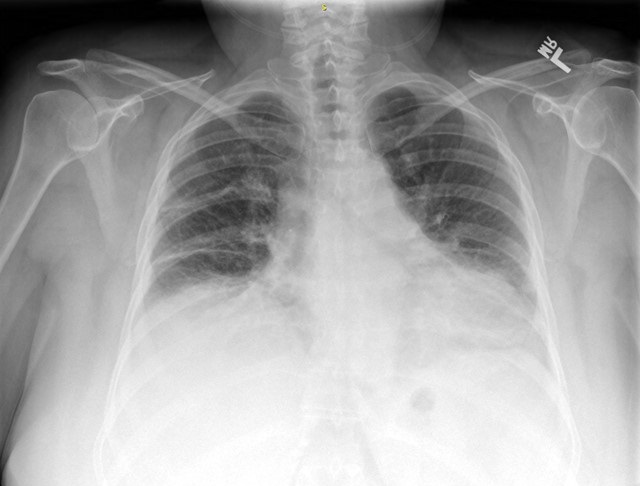
March 2019 Pulmonary Case of the Month: A 59-Year-Old Woman with Fatigue

Lewis J. Wesselius, MD1

Michael B. Gotway, MD2

1Department of Pulmonary Medicine and 2Department of Radiology

Mayo Clinic Arizona

Scottsdale, AZ USA


History of Present Illness

A 59-year-old woman from Kingman, Arizona had a one-year history of fatigue with some shortness of breath. For this reason, she saw her primary care physician.

Past Medical History, Social History and Family History

She has no significant past medical history. She does not smoke. Family history is noncontributory.

Physical Examination

Physical examination was unremarkable.

Which of the following should be done? (Click on the correct answer to be directed to the second of seven pages)

  1. Chest x-ray
  2. Complete blood count
  3. Electrolytes, blood urea nitrogen and creatinine
  4. Liver panel
  5. All of the above

Cite as: Wesselius LJ, Gotway MB. March 2019 pulmonary case of the month: A 59-year-old woman with fatigue. Southwest J Pulm Crit Care. 2019;18(3):52-7. doi: https://doi.org/10.13175/swjpcc008-19 PDF 

Thursday
Jan242019

Co-Infection with Nocardia and Mycobacterium Avium Complex (MAC) in a Patient with Acquired Immunodeficiency Syndrome 

Zahira Babwani DO

Kenneth Wojnowski Jr DO

Sunil Kumar MD

Broward Health Medical Center

Fort Lauderdale, FL USA

Abstract

We present a case in which a patient with acquired immunodeficiency syndrome (AIDS) and nocardiosis was found to have co-infection with Mycobacterium avium complex (MAC). Despite the fact that MAC is a known colonizer of the pulmonary system, ​ it is possible to have co-infection and a high degree of suspicion is necessary to ensure prompt treatment of both organisms. We wish to describe how radiologic findings were instrumental in guiding our differential diagnosis.

Case Report

History of Present Illness: A 64-year-old man with history of alcohol and tobacco abuse presented with a chronic, productive cough for 5-6 months. Associated symptoms included shortness of breath and 30-pound weight loss. He denied all other symptoms.

Physical Exam: Pertinent positives revealed temporal wasting, poor dental hygiene, oral thrush and diffuse rhonchi bilaterally. Initial vital signs were within normal limits.

Laboratory and Radiology: Pertinent laboratory findings revealed leukocytosis with a left shift. Viral respiratory polymerase chain reaction (PCR) testing was negative. Human immunodeficiency virus (HIV) testing was positive with a CD4 count of 46 cells/mm3. QuantiFERON gold testing was negative. Sputum cultures, acid-fast bacilli (AFB) and blood cultures were obtained. Bronchoalveolar lavage (BAL) was performed with no evidence of Pneumocystis jirovecii (PJP). Chest X-ray (CXR) and computed tomography (CT) of the chest (Figure 1) revealed a multifocal right lung abscess with complex pleural fluid, empyema, nodular cavitary lesion in the left lower lobe and hilar lymphadenopathy.

Figure 1. Panel A: initial chest X-ray shows a complex infiltrate and effusion in the right lung. There is a cavitary lesion with air-fluid level vs lung abscess on the right. A nodule or consolidation is present in the left lung base. Panel B: A representative image from the initial CT of the chest showing a multifocal right lung abscess and complex pleural fluid.

Hospital Course: ​After admission, the patient was started on broad spectrum antimicrobials with vancomycin and piperacillin-tazobactam. A thoracentesis was performed due to right sided pleural effusion which yielded 65 cc of thick, purulent, green fluid. Thoracotomy with complete decortication of the right lung was performed with biopsies of the abscesses. Two 32-French chest tubes were placed due to the presence of multiple intraparenchymal lung abscesses, loculations, and empyema. Biopsy and pleural fluid cultures grew gram positive, beaded organisms which were later identified as nocardia, with no evidence of MAC or Mycobacterium tuberculosis (MTB). The patient was started on amikacin, meropenem and trimethoprim-sulfamethoxazole for newly diagnosed pulmonary nocardiosis. MAC prophylaxis was initiated due to his low CD4 count. After initiation of therapy for nocardiosis, three sputum AFB cultures began to stain positive. Since nocardiosis stains weakly positive for AFB, we initially did not suspect non-tuberculous Mycobacteria (NTM). Repeat CT scan of the chest (Figure 2) revealed ground glass opacities, nodular densities and both mediastinal and hilar lymphadenopathy.

Figure 2. Panel A: after initiation of treatment for nocardiosis, improvement of right empyema and cavitary lesion with bilateral patchy airspace disease right greater than left. Panel B: CT of the chest after initiation of treatment for nocardiosis, prominent lymph nodes in the hilar regions and mediastinum. less cavitation than the previous study. There are innumerable ground glass and nodular densities throughout both lungs, right greater than left.

Suspicion for active MAC co-infection was raised, the prophylactic dose of azithromycin was increased to the treatment dose, and ethambutol was initiated. After three weeks of intravenous amikacin, meropenem and trimethoprim-sulfamethoxazole the patient showed considerable improvement in his respiratory symptoms and was transitioned to oral trimethoprim-sulfamethoxazole for outpatient treatment of nocardiosis with continuation of ethambutol and clarithromycin for MAC.

Discussion

The Mycobacterium Avium Complex ​(MAC) is a Non-tuberculous mycobacterium (NTM) that is commonly found in patients with HIV and a CD4 count of less than 50. The diagnosis of NTM is challenging due to the fact that the organism is a known colonizer of the pulmonary system (1) ​. Supportive radiologic evidence is needed to distinguish colonization from active infection (2).

Common CT findings of nocardiosis include ground glass opacities, lung nodules, cavitation, pleural effusion and masses (3)​. The presence of mediastinal and hilar lymphadenopathy is the most common finding in immunosuppressed patients with MAC infection but is not​ a usual feature of pulmonary nocardiosis (3,4) ​. Our​ patient’s repeat CT scan showed mediastinal and hilar lymphadenopathy with improvement of cavitary lesions which suggests improvement of CT findings related to nocardiosis, but persistent findings related to NTM (5). This led us to believe that the patient was appropriately treated for nocardiosis, but with an underlying presence of active MAC infection that presented with atypical radiographic findings. As per the American Thoracic Society (ATS) guidelines for NTM pulmonary infection (6)​ ​, this patient’s pulmonary symptoms, radiological evidence on the chest CT, and positive AFB cultures from at least two separate expectorated sputum samples lends credibility to MAC as a true active infection in the setting of nocardiosis and AIDS. The patient was appropriately placed on clarithromycin and ethambutol as an outpatient, and our suspicions were confirmed for MAC with no evidence of MTB by PCR testing 5 weeks after initial AFB smears were collected.

Co-infection with Nocardiosis and MAC may be underestimated since they both often develop in immunocompromised hosts. MAC, along with other NTM species account for 20% of mycobacterium pulmonary infections in HIV infected patients (5)​. Nocardia accounts for less than 3% of pulmonary infections in HIV infected patients (5)​. A high degree of clinical suspicion is imperative to promptly treat infection with both organisms.

References

  1. Young J, Balagopal A, Reddy NS, Schlesinger LS. Differentiating colonization from infection can be difficult Nontuberculous mycobacterial infections: Diagnosis and treatment. Patient Care. 2007. Available at: http://www.patientcareonline.com/infection/differentiating-colonization-infection-can-be-difficult-nontuberculous-mycobacterial-infections (accessed 10/3/18).
  2. Trinidad JM, Teira R, Zubero S, Santamaría JM.Coinfection by Nocardia asteroides and Mycobacterium avium- intracellulare in a patient with AIDS. Enferm Infecc Microbiol Clin. 1992 Dec;10(10):630-1. [PubMed]
  3. Kanne JP, Yandow DR, Mohammed TL, Meyer CA. CT findings of pulmonary nocardiosis. AJR Am J Roentgenol. 2011 Aug;197(2):W266-72. [CrossRef] [PubMed]
  4. Erasmus JJ, McAdams HP, Farrell MA, Patz EF Jr. Pulmonary nontuberculous mycobacterial infection: radiologic manifestations. Radiographics. 1999 Nov-Dec;19(6):1487-505. [PubMed]
  5. Benito N, Moreno A, Miro JM, Torres A. Pulmonary infections in HIV-infected patients: an update in the 21st century. Eur Respir J. 2012 Mar;39(3):730-45. [CrossRef] [PubMed]
  6. Griffith DE, Aksamit T, Brown-Elliott BA, et al. An official ATS/IDSA statement: diagnosis, treatment, and prevention of nontuberculous mycobacterial diseases. Am J Respir Crit Care Med. 2007 Feb 15;175(4):367-416. [CrossRef] [PubMed]

Cite as: Babwani Z, Wojnowski K Jr, Kumar S. Co-Infection with Nocardia and Mycobacterium avium complex (MAC) in a patient with acquired immunodeficiency syndrome. Southwest J Pulm Crit Care. 2019;18(1):22-5. doi: https://doi.org/10.13175/swjpcc123-18 PDF

Tuesday
Jan082019

Progressive Massive Fibrosis in Workers Outside the Coal Industry: A Case Series from New Mexico

Landon Casaus, MD1

Sapna Bhatia, MD1

Akshay Sood, MD, MPH1, 2


1Department of Internal Medicine

University of New Mexico School of Medicine

Albuquerque, NM, USA

2Miners’ Colfax Medical Center

Raton, NM USA


Abstract

Four clinical patterns of diffuse lung disease may be seen with silicosis: acute silicosis or silicoproteinosis (the latter resembling pulmonary alveolar proteinosis), simple nodular sclerosis, accelerated silicosis, and progressive massive fibrosis (PMF). The intensity and duration of exposure as well as host susceptibility dictates the presentation and progression of PMF. Although most cases of PMF in the literature are reported among coal miners in whom this disease has shown a recent increase in prevalence, this disease can also be seen in exposed workers outside the coal industry. In this article, we will review the clinical, physiological, and pathological manifestations of the disease, illustrated by three case examples of PMF among non-coal miners from New Mexico. Diagnosis and management of patients with PMF can be difficult, and carries medicolegal implications for the patient. Physicians and policymakers need to be aware of PMF in workers exposed to silica within and outside the coal industry.

Introduction

The worldwide prevalence of silicosis peaked by the beginning of the 20th century during the development of mechanized industry (1). Outbreaks of silicosis are still noted in the developed world, particularly where workers are consistently exposed to silica particles that are small enough to be inhaled (≤10 µm in diameter) and at levels above a “safe” concentration (action level of 25 µm/m3 as a time-weighted average over an 8-hour work day, as recommended by the U.S. Occupational Safety and Health Administration or OSHA) (2,3). The four Appalachian coal mining states of Pennsylvania, West Virginia, Virginia, and Kentucky accounted for more than 75 percent of all silicosis-related deaths in the United States (U.S.) in 2007 (4).  A recent study however indicates that the age-standardized mortality rate from silicosis in the U.S. in 2014 was amongst the highest in the mining intense regions of the Southwest, particularly in the Four Corners area where the borders of New Mexico, Arizona, Utah, and Colorado meet (5). The number of diagnosed silicosis cases has increased in New Mexico between 2000 and 2011, and residents of New Mexico are twice as likely to die from or with silicosis when compared to the rest of the country for reasons that are unexplained (6).

Four clinical patterns of diffuse lung disease may be seen with silicosis: acute silicosis or silicoproteinosis (the latter resembling pulmonary alveolar proteinosis), simple nodular sclerosis, accelerated silicosis, and progressive massive fibrosis (PMF). PMF represents the coalescence of multiple small pneumoconiotic opacities to form larger opacities or conglomerate masses measuring over 10 millimeters in size on a chest radiograph, with smaller rounded opacities usually seen in simple silicosis. Silicotic opacities are classified on their shape, size, and profusion using the International Labour Organization’s (ILO) International Classification of Radiographs for Pneumoconiosis system (commonly referred to as B reads) (7-9). The 1970-2017 radiographic data from the National Institute for Occupational Safety and Health (NIOSH) surveillance program concluded that the national prevalence of coal workers’ pneumoconiosis in coal miners with 25 years or more of tenure now exceeds 10% (10). This is an increase from the previous estimate of 7% in 2012 (11,12). A resurgence of progressive massive fibrosis in coal miners has also been described, particularly those working in smaller mines (13). The rate of PMF in silica exposed workers outside of the coal mine industry, similar to those illustrated in this paper, is unknown. We herein describe three New Mexico non-coal miners with PMF that were followed at the University of New Mexico Occupational Pulmonary Medicine Clinic. Each of the three cases had already received compensation under the United States Energy Employees Occupational Illness Compensation Program, based upon prior abnormal B reads of chest radiographs. The epidemiology, pathogenesis, and management of PMF is also reviewed.

Case reports

Case 1

An 83-year-old man presented in 2017 with worsening dyspnea over the prior 10 years. He worked at a federal national laboratory in northern New Mexico, from 1962-1992 as a construction worker. His work included digging ditches, removing insulation, demolishing buildings, breaking up concrete with jackhammers, and working around sandblasters in enclosed areas, without any respiratory protection. He had a 5-pack year smoking history, and quit 50 years prior.

A 2017 chest radiograph showed small, upper lobe predominant, nodular opacities. A high-resolution computed tomography (CT) scan in 2009 showed innumerable micronodules in the upper lobes of the lung with a centrilobular distribution.  A repeat CT scan obtained in 2017 (Figure 1) showed new-onset coalescence of several upper lobe nodules, as large as 1.5 cm x 2 cm.

Figure 1. Computed tomography scan of the chest showing several silicotic opacities in both lung apices and coalescence to progressive massive fibrosis in right apex.

His pulmonary function tests (PFT) showed mild obstruction with evidence of air trapping. A diagnostic bronchoscopy showed no evidence of infection or neoplasm.

Case 2

A 78-year-old man presented in 2014 with several-years history of progressive New York Heart Association Class III dyspnea. The patient worked as an underground uranium miner from 1960 to 1989 where he was exposed to hauling, “mucking” (a term referring to the loading of fragmented ore), and blasting. He wore a respirator intermittently. He had a five-pack year smoking history, quitting in 1981.

Chest x-ray showed innumerable micronodules, predominately in the upper lobes.  A CT scan of the chest with 3 mm cuts in 2012 showed innumerable upper lobe predominant micronodules in a perilymphatic and centrilobular distribution, with coalescence in the upper lobes. Repeat CT scans in 2014 and 2015 demonstrated no disease progression (Figure 2).

Figure 2. Computed tomography scan of the chest demonstrating progressive massive fibrosis in the right upper lung and several silicotic opacities in bilateral upper lungs.

PFTs showed a mild restrictive defect. An infectious etiology was ruled out by negative sputum acid fast bacilli (AFB), and bacterial smears and cultures.

Case 3

A 79-year-old man presented with dyspnea at rest and upon exertion, and chronic bronchitis symptoms, with occasional hemoptysis. The patient worked as an underground uranium miner from 1959-1980 performing drilling, blasting and “mucking”, with significant self-reported exposure to dust and without use of respiratory protection. The patient reported a 15-pack year smoking history, but quit in 1976.

A chest x-ray showed hilar and mediastinal nodal calcifications with small scattered lung nodules. A HRCT scan of the chest in 2016 (Figure 3) showed multiple calcified nodules as well as calcified hilar and mediastinal lymph nodes.

Figure 3. Computed tomography scan of the chest demonstrating progressive massive fibrosis with evidence of traction in both lobes.

Conglomerate masses in the upper lobes measuring up to 3.3 cm were noted and moderate background emphysematous changes were also noted.  The PFTs on initial evaluation were within normal limits. He was noted to be hypoxic on room air, necessitating 2 L/min oxygen supplementation. Sputum AFB smears and cultures were negative.

Discussion

PMF is seen in workers employed in industries that cut, grind, or drill silica-containing materials such as concrete, masonry, tile and/or rock (3). Most cases of PMF in the literature have been reported among coal miners, likely a reflection of the fact that coal miners undergo active surveillance due to governmental regulations (12). Although more commonly believed to occur in underground coal miners, PMF can be seen in surface coal miners as well (14). PMF outside the coal industry has been described in limited studies of barium miners, sandblasters, blacksmiths, welders, metal polishers, and quartz surface fabricators (15-17). More recently, PMF has been reported in ‘distressed’ denim jean industry workers (18). In this case series, we report PMF in New Mexico construction and uranium workers.

The latency for PMF is usually 10-30 years. Latency is greatly impacted by the exposure concentration and duration, as well as type of silica exposure. Additionally, it is influenced by underlying diseases, genetics, and smoking. Although PMF typically occurs in a setting of high cumulative dust exposures (14), some studies indicate that the host patterns of deposition and clearance of dust may be more relevant (19).

The pathogenesis of PMF is not completely understood; however, it is known that alveolar macrophages initiate a complex cascade results in inflammation and fibrosis (20). Histopathological findings include nodules, usually located near the respiratory bronchioles, composed of silica particles surrounded by whorled collagen in concentric layers. Larger masses of collagen define the lesion of PMF, which may be associated with avascular necrosis in the center and endarteritis in the periphery (21). The extensive fibrotic reaction in PMF is associated with high serum levels of interleukin (IL)-8 and intercellular adhesion molecule (ICAM)-1, which are important as neutrophil attractants and adhesion molecules (22).

The clinical diagnosis of PMF has three requirements: the patient must have a history of inhalational silica exposure significant enough to cause disease; chest imaging must be consistent with PMF; and other illnesses that mimic PMF must be reasonably ruled out (1). The disease presentation of PMF is highly variable. Patients may have debilitating symptoms of dyspnea on exertion and exercise intolerance, obstructive and/or restrictive patterns on PFTs, as well as experience complications such as cor pulmonale, spontaneous pneumothorax, and hypoxic respiratory failure (23). On the other hand, a normal spirogram is described in up to 11% of subjects with PMF, as also noted in Case 3 above (23).  The level of pulmonary impairment in patients with PMF generally increases with increasing radiologic size of large opacities (23). Spirometry is repeated upon follow-up visits to assess for functional deterioration (24). Invasive tests such as arterial blood gas or cardiopulmonary exercise test are usually not indicated. Surveillance chest radiographs are classified for small and large opacities using the International Labour Organization’s (ILO) International Classification of Radiographs for Pneumoconiosis system (7-9). CT scan of the chest is more sensitive in diagnosing PMF than chest radiographs, and may be considered if the radiograph fails to show large opacities but demonstrates small opacities of relatively larger diameter or a tendency for opacities to coalesce (25,26). Lung tissue for histology or mineral analysis is rarely needed. The presence of atypical features in a patient with simple silicosis such as fever, hemoptysis, worsening dyspnea, weight loss, disproportionate fatigue, and the presence of a new infiltrate or cavitation of a pre-existing lesion on chest imaging should prompt the clinician to look for PMF, tuberculosis, or lung cancer. Patients with PMF are at elevated risk for concomitant tuberculosis. This risk is directly proportional to the level of profusion of silicotic small opacities (27), and the risk in patients with the highest level of profusion is comparable to that in patients with HIV infection (3). Autopsy studies from Welsh coal workers during the period 1952–1954 demonstrated tubercle bacilli in as many as 35% of cases with PMF (21). A recently published study from Brazil reported coexisting microbiologically confirmed tuberculosis in about half of patients with PMF, raising concerns about tuberculosis infection as a risk factor for the development of PMF 15. Patients with silicosis are also at high risk for lung cancer (28), with a greater risk for lung cancer described in patients with PMF as compared to patients with simple coal workers pneumoconiosis in one study (29). Positron emission tomography with F-18 fluorodeoxyglucose is of limited utility in differentiating malignancy from PMF lesions (30).

The prevention of PMF remains a focus at the exposed workplace. This includes primary prevention such as worker education; control of airborne dust exposure via engineering and work practice interventions such as improving ventilation, providing a means of exhaust, adding water to the cutting surface, and using enclosed cabs or booths; and use of respiratory protective devices (3). In June 2018, OSHA mandated personal breathing zone air sampling to monitor exposure and medical surveillance of workers with exposure above the permissible exposure limits (31). Medical surveillance constitutes secondary prevention, facilitating early diagnosis and treatment. Surveillance should be done periodically and should include a medical examination and occupational questionnaire, chest radiograph with B read interpretation, tuberculosis screening, and spirometry, with referral of affected workers to a pulmonologist or occupational medicine physician for further evaluation (32).

Once PMF has been diagnosed, it is important to immunize the patient against influenza and pneumococcal infection, assess the need for oxygen supplementation, and encourage pulmonary rehabilitation. Exclusion of active tuberculosis is recommended and screening for latent tuberculosis infection by either skin testing or interferon gamma release assay should be considered (33). Systemic corticosteroids, inhaled aluminum citrate, poly(vinlypyridine-N-oxide) and whole lung lavage are unlikely to benefit patients with PMF and lung transplantation may be considered (4). 

Patients with PMF are considered ‘totally disabled’ from coal mine employment under the Black Lung Benefits Act in the United States. Outside the coal industry, they may be eligible for benefits under the Social Security Impairment system or the state workers’ compensation systems.

Conclusion

PMF represents the coalescence of smaller radiographic pneumoconiotic opacities to those over 10 millimeters in size. The rate of PMF in American coal miners has recently increased. Although most cases of PMF are reported among coal miners, this is likely a reflection of the fact that coal miners undergo active surveillance due to governmental regulations 12. In this case series, we report PMF in workers outside the coal industry. Physicians and policymakers need to be aware of this condition in workers exposed to silica within and outside the coal industry.

Acknowledgments

Guarantor: Landon Casaus, M.D., takes responsibility for the content of the manuscript, including the data and analysis.

Author contributions: All authors had full access to all the data in the study and take responsibility for the integrity of the data and the accuracy of the data analysis. All authors contributed substantially to the data analysis and interpretation and the writing of the manuscript.

Financial/non-financial disclosures: All authors report no conflict of interest.

Abbreviations List

  • AFB: Acid fast bacilli
  • CT: computed tomography
  • HIV: Human immunodeficiency virus
  • HRCT: High resolution computed tomography
  • IL: interleukin
  • ICAM: Intercellular adhesion molecule
  • NIOSH: National Institute for Occupational Safety and Health
  • OSHA: Occupational Safety and Health Administration
  • PMF: Progressive massive fibrosis
  • PFT: Pulmonary function test
  • US: United States

References

  1. Banks D. Silicosis. In: Rosenstock L, Cullen M, Brodkin C, Redlich C, eds. Textbook of clinical occupational and environmental medicine. 2nd edition ed. China: Elsevier Saunders; 2005.
  2. Occupational Safety and Health Administration. Occupational Exposure to Respirable Crystalline Silica; Final Rule. In. Federal Register. Vol 81. Washington DC: Government Publishing Office; 2016:16285-16890. [PubMed]
  3. Adverse effects of crystalline silica exposure. American Thoracic Society Committee of the Scientific Assembly on Environmental and Occupational Health. Am J Respir Crit Care Med. 1997 Feb;155(2):761-8. [CrossRef] [PubMed]
  4. National Institute for Occupational Safety and Health, Department of Health and Human Services Centers for Disease Control and Prevention. The Work-Related Lung Disease Surveillance Report 2007. Available at: https://www.cdc.gov/niosh/docs/2008-143/default.html.
  5. Dwyer-Lindgren L, Bertozzi-Villa A, Stubbs RW, Morozoff C, Shirude S, Naghavi M, Mokdad AH, Murray CJL. Trends and Patterns of Differences in Chronic Respiratory Disease Mortality Among US Counties, 1980-2014. JAMA. 2017;318(12):1136-49. [CrossRef] [PubMed]
  6. New Mexico Occupational Health Surveillance Program New Mexico Department of Health. Silicosis in New Mexico Infographic. Available at https://nmhealth.org/about/erd/eheb/ohsp/.
  7. Welch LS, Hunting KL, Balmes J, Bresnitz EA, Guidotti TL, Lockey JE, Myo-Lwin T. Variability in the classification of radiographs using the 1980 International Labor Organization Classification for Pneumoconioses. Chest. 1998 Dec;114(6):1740-8. [CrossRef] [PubMed]
  8. Halldin CN, Blackley DJ, Petsonk EL, Laney AS. Pneumoconioses Radiographs in a Large Population of U.S. Coal Workers: Variability in A Reader and B Reader Classifications by Using the International Labour Office Classification. Radiology. 2017 Sep;284(3):870-6. [CrossRef] [PubMed]
  9. International Labor Organization. Guidelines for the use of the ILO International Classification of Radiographs of Pneumoconioses. In. Vol Occupational Safety and Health Series, No. 22. 2000 ed. Geneva: International Labor Office; 2002.
  10. Blackley DJ, Halldin CN, Laney AS. Continued increase in prevalence of coal workers' pneumoconiosis in the United States, 1970-2017. Am J Public Health. 2018 Sep;108(9):1220-2. [CrossRef] [PubMed]
  11. Laney AS, Weissman DN. Respiratory diseases caused by coal mine dust. J Occup Environ Med. 2014 Oct;56 Suppl 10:S18-22. [CrossRef] [PubMed]
  12. Blackley DJ, Halldin CN, Laney AS. Resurgence of a debilitating and entirely preventable respiratory disease among working coal miners. Am J Respir Crit Care Med. 2014 Sep 15;190(6):708-9. [CrossRef] [PubMed]
  13. Blackley DJ, Halldin CN, Wang ML, Laney AS. Small mine size is associated with lung function abnormality and pneumoconiosis among underground coal miners in Kentucky, Virginia and West Virginia. Occup Environ Med. 2014 Oct;71(10):690-4. [CrossRef] [PubMed]
  14. Halldin CN, Reed WR, Joy GJ, et al. Debilitating lung disease among surface coal miners with no underground mining tenure. J Occup Environ Med. 2015 Jan;57(1):62-7. [CrossRef] [PubMed]
  15. Ferreira AS, Moreira VB, Ricardo HM, Coutinho R, Gabetto JM, Marchiori E. Progressive massive fibrosis in silica-exposed workers. High-resolution computed tomography findings. J Bras Pneumol. 2006 Nov-Dec;32(6):523-8. [CrossRef] [PubMed]
  16. Friedman GK, Harrison R, Bojes H, Worthington K, Filios M; Centers for Disease Control and Prevention (CDC). Notes from the field: silicosis in a countertop fabricator - Texas, 2014. MMWR Morb Mortal Wkly Rep. 2015 Feb 13;64(5):129-30. [PubMed]
  17. Seaton A, Ruckley VA, Addison J, Brown WR. Silicosis in barium miners. Thorax. 1986;41(8):591-5. [CrossRef] [PubMed]
  18. Bakan ND, Özkan G, Çamsari G, Gür A, Bayram M, Açikmeşe B, Çetinkaya E. Silicosis in denim sandblasters. Chest. 2011;140(5):1300-4. [CrossRef] [PubMed]
  19. Douglas AN, Robertson A, Chapman JS, Ruckley VA. Dust exposure, dust recovered from the lung, and associated pathology in a group of British coalminers. Br J Ind Med. 1986 Dec;43(12):795-801. [CrossRef] [PubMed]
  20. Sayan M, Mossman BT. The NLRP3 inflammasome in pathogenic particle and fibre-associated lung inflammation and diseases. Part Fibre Toxicol. 2016 Sep 20;13(1):51. [CrossRef] [PubMed]
  21. Davies DG, James WR, Rivers D, Thomson S. The prevalence of tuberculosis at necropsy in progressive massive fibrosis of coalworkers. Br J Ind Med. 1957 Jan;14(1):39-42. [PubMed]
  22. Lee JS, Shin JH, Choi BS. Serum levels of IL-8 and ICAM-1 as biomarkers for progressive massive fibrosis in coal workers' pneumoconiosis. J Korean Med Sci. 2015 Feb;30(2):140-4. [CrossRef] [PubMed]
  23. Yeoh CI, Yang SC. Pulmonary function impairment in pneumoconiotic patients with progressive massive fibrosis. Chang Gung Med J. 2002 Feb;25(2):72-80. [PubMed]
  24. Fernandez Alvarez R, Martinez Gonzalez C, Quero Martinez A, Blanco Perez JJ, Carazo Fernandez L, Prieto Fernandez A. Guidelines for the diagnosis and monitoring of silicosis. Arch Bronconeumol. 2015;51(2):86-93. [CrossRef] [PubMed]
  25. Martinez Gonzalez C, Fernandez Rego G, Jimenez Fernandez-Blanco JR. [Value of computerized tomography in the diagnosis of complicated pneumoconiosis in coal miners]. Arch Bronconeumol. 1997;33(1):12-5. [CrossRef]
  26. Ooi GC, Tsang KW, Cheung TF, Khong PL, Ho IW, Ip MS, Tam CM, Ngan H, Lam WK, Chan FL, Chan-Yeung M. Silicosis in 76 men: qualitative and quantitative CT evaluation-clinical-radiologic correlation study. Radiology. 2003;228(3):816-25. [CrossRef] [PubMed]
  27. Rees D, Murray J. Silica, silicosis and tuberculosis. Int J Tuberc Lung Dis. 2007 May;11(5):474-84. [PubMed]
  28. Poinen-Rughooputh S, Rughooputh MS, Guo Y, Rong Y, Chen W. Occupational exposure to silica dust and risk of lung cancer: an updated meta-analysis of epidemiological studies. BMC Public Health. 2016 Nov 4;16(1):1137. [CrossRef] [PubMed]
  29. Tomaskova H, Jirak Z, Splichalova A, Urban P. Cancer incidence in Czech black coal miners in association with coalworkers' pneumoconiosis. Int J Occup Med Environ Health. 2012 Jun;25(2):137-44. [CrossRef] [PubMed]
  30. Reichert M, Bensadoun ES. PET imaging in patients with coal workers pneumoconiosis and suspected malignancy. J Thorac Oncol. 2009 May;4(5):649-51. [CrossRef] [PubMed]
  31. Occupational Safety and Health Administration. Respirable crystalline silica. In. Vol 1926. Washington DC: Occupational Safety and Health Administration; Federal Register; 2016:1153. Available from: https://www.osha.gov/silica/SilicaConstructionRegText.pdf  (last accessed 2018 Jul 1125).
  32. Deslauriers JR, Redlich CA. Silica Exposure, Silicosis, and the New Occupational Safety and Health Administration Silica Standard. What Pulmonologists Need to Know. Ann Am Thorac Soc. 2018 Dec;15(12):1391-1392. [CrossRef] [PubMed]
  33. Lewinsohn DM, Leonard MK, LoBue PA, et al. Official American Thoracic Society/Infectious Diseases Society of America/Centers for Disease Control and Prevention Clinical Practice Guidelines: Diagnosis of Tuberculosis in Adults and Children. Clin Infect Dis. 2017 Jan 15;64(2):111-5. [CrossRef] [PubMed] 

Cite as: Casaus L, Bhatia S, Sood A. Progressive massive fibrosis in workers outside the coal industry: A case series from New Mexico. Southwest J Pulm Crit Care. 2019;18(1):10-9. doi: https://doi.org/10.13175/swjpcc110-18 PDF 

Saturday
Dec012018

December 2018 Pulmonary Case of the Month: A Young Man with Multiple Lung Masses

Lewis J. Wesselius, MD

Department of Pulmonary Medicine

Mayo Clinic Arizona

Scottsdale, AZ USA


History of Present Illness

A 28-year-old man from Tennessee has been feeling ill with malaise and weight loss for the past 3 months. He had been in the in the Palm Springs area a few weeks prior to becoming ill. He works as a musician.

Past Medical History, Social History and Family History

He has a history of Wolf-Parkinson-White syndrome and had a prior ablation procedure at age 16. He does not smoke tobacco but does smoke marijuana occasionally. Family history is noncontributory.

Physical Examination

Physical examination was unremarkable.

Which of the following are indicated at this time? (Click on the correct answer to be directed to the second of eight pages)

  1. Bronchoscopy with EBUS
  2. Chest X-ray
  3. VATS
  4. 1 and 3
  5. All of the above

Cite as: Wesselius LJ. December 2018 pulmonary case of the month: a young man with multiple lung masses. Southwest J Pulm Crit Care. 2018;17(6):138-45. doi: https://doi.org/10.13175/swjpcc118-18 PDF 

Friday
Sep212018

Antibiotics as Anti-inflammatories in Pulmonary Diseases

Richard A. Robbins, MD

Phoenix Pulmonary and Critical Care Research and Education Foundation

Gilbert, AZ USA


Abstract

The currently available evidence for the use of chronic antibiotic therapy, principally macrolides and tetracyclines, as anti-inflammatory therapy in pulmonary disorders is reviewed. Historically, treatment of a number of chronic diseases with tetracyclines showed modest benefits but reports of the successful treatment of diffuse panbronchiolitis with erythromycin stimulated research in other lung diseases as well as shifting the focus from tetracyclines to macrolides. Chronic macrolide therapy is now recommended for patients with frequent exacerbations of cystic fibrosis and COPD and considerable evidence exists for potential benefits in asthma. There is also evidence of macrolide efficacy in the prevention of obliterative bronchiolitis after lung transplantation. Small trials have suggested possible benefit of macrolides in IPF. Taken together these suggest a potential for antibiotics, particularly macrolides, in some pulmonary inflammatory disorders.

History

Based on responses to antibiotics the concept arose over 70 years ago that several common diseases might have an infectious origin. In 1949, Thomas McPherson Brown reported favorable results of tetracycline treatment for rheumatoid arthritis patients at the 7th International Congress on Rheumatic Diseases (1). It was hypothesized these effects were due to a mycoplasma infection. However, the beneficial effects of cortisone in the treatment of arthritis were described at the same meeting. The effect of tetracycline paled beside that of steroids, and the salutary effects of antibiotics on rheumatoid arthritis were largely ignored.

Acne rosacea is a common, chronic dermatologic condition, whose cause remains unknown. Tetracyclines were the first systemic drugs used in the treatment of rosacea, and have been the mainstay therapy for more than 50 years (2). More recently, sub-antimicrobial doses of tetracyclines have been shown to be effective in rosacea presumably through an anti-inflammatory effect (3). Dermatitis herpetiformis is a disease now thought to be secondary to gluten sensitivity. However, this disorder has been treated with dapsone for over 60 years despite its non-infectious origin (4).

Tetracyclines have long been used for periodontal disease with clinical benefit presumed to be from their antimicrobial properties. However, as early as 1983, Golub (5) proposed that tetracyclines might have a beneficial effect by modifying inflammation. Now the tetracyclines are thought to exert their beneficial effects by anti-inflammatory effects, anti-collagenase effects, and a reduction in bone loss (6).

In 1959 the late Neil Cherniack published a double-blind study of 67 patients with chronic bronchitis or bronchiectasis treated with tetracycline, penicillin, a combination of oleandomycin and penicillin, or placebo for 3 to 22 months (7). Patients who received tetracycline had significantly fewer lower respiratory illnesses than those treated with placebos or penicillin. The average duration of these illnesses was also shorter in patients treated with tetracycline.

The anti-inflammatory effects of the macrolides were brought to light because of their effects on an uncommon pulmonary disease, diffuse panbronchiolitis (DPB). DPB is a rare disease seen in Japan and characterized by a chronic inflammatory neutrophilic inflammation of the airways, DPB has a 5-year survival rate of just 63% but only 8% when patients’ airways became colonized with Pseudomonas aeruginosa (8). However, in the early 1980s it was discovered that chronic treatment with erythromycin resulted in dramatically improved 5-year survival to 92% (8). This improvement occurred despite a failure to eliminate the bacterial colonization and was associated with a dramatic decrease in the accumulation of airway neutrophils (8,9). Interestingly, the effect on neutrophilic inflammation was found to be a nonspecific effect of the macrolides. Other macrolides (clarithromycin, roxithromycin and azithromycin) produced a similar suppression of the neutrophilic inflammation (10).

Gradually, with a better understanding of the pathogenesis of these common disease and basic studies examining anti-inflammatory effects, the macrolides and tetracyclines were recognized as anti-inflammatories. Inflammation is proposed to play a role in the pathogenesis of a number of pulmonary disorders. The encouraging results of the above suggested that macrolides and tetracyclines might be beneficial in pulmonary inflammatory conditions. Studies have examined a number of disorders including cystic fibrosis, chronic obstructive pulmonary disease, bronchiectasis, and asthma.

Anti-inflammatory Mechanisms of Action

Macrolides and tetracyclines exert their antibacterial effects by inhibiting bacterial protein synthesis. Although the anti-inflammatory mechanisms of action of the tetracyclines and macrolides are likely multiple, one important mechanism by both is a reduction in production of a multitude of pro-inflammatory cytokines. Most of these cytokines are regulated at the transcriptional level through proteins such as nuclear factor-κβ (NF- κβ), activator protein-1 (AP-1) and/or p38 mitogen-activated protein kinases (p38 MAPK). Although the studies have varied depending on the in vitro systems examined, most have described a shortening of the half-life of pro-inflammatory cytokine mRNA usually through effect on one or more of the transcriptional control proteins (10-13).

Cystic Fibrosis

A major step in the use of antibiotics as anti-inflammatories occurred with the introduction of macrolides as adjunctive therapy in cystic fibrosis in 2003. Like diffuse panbronchiolitis, airways of cystic fibrosis patients show chronic inflammation with neutrophils which are often infected with Pseudomonas aeruginosa. Saiman et al. (14) conducted a multicenter, randomized, double-blind, placebo-controlled trial of azithromycin in cystic fibrosis patients infected with Pseudomonas. They found a reduction in exacerbations and greater weight gain in those treated with azithromycin compared to control. Following several confirming studies, cystic fibrosis patients are now commonly treated with macrolide antibiotics, especially when infected with Pseudomonas (15).

Tetracyclines have been less commonly used probably because of the staining of teeth and bone in younger, growing children. However, a recent small trial of 19 adult cystic fibrosis treated with chronic doxycycline showed an improvement in FEV1 and an increase in time to the next exacerbation compared to 20 placebo-treated controls (16). This might suggest an alternative in older patients or those at high risk for side effects from macrolides.

Non-CF Bronchiectasis

Long-term treatment with antibiotics has been recommended in patients with bronchiectasis and frequent exacerbations (17). This is based on studies showing decreased rates of exacerbations and some improvement in quality of life. It is not clear whether this effect is due to the antibacterial or anti-inflammatory properties of macrolides. In addition to Cherniak’s tetracycline trial which included bronchiectatics (7), an early MRC trial in 1957 showed that long-term twice weekly oxytetracycline over 1 year led to reduced sputum purulence, fewer days confined to bed and fewer days off work (18). Later trials in non-CF bronchiectasis have been done primarily with azithromycin and it is noted that there is an increased risk of macrolide-resistant organisms developing in these patients, as well as other risks associated with macrolide therapy including ototoxicity and QT prolongation (19).

Chronic Obstructive Pulmonary Disease

Chronic obstructive pulmonary disease (COPD) is one of the most expensive diseases to treat (20). A number of studies examining costs of COPD have shown that exacerbations, especially those resulting in hospitalization, account for the majority of costs (21,22). Although treatment with glucocorticoids, long-acting beta2-agonists, and long-acting muscarinic antagonists reduce the frequency of acute exacerbations, COPD patients receiving all three of these medications still average 1.4 acute exacerbations per year (23). Beginning in the early 2000’s there were a number of studies that reported an improvement in COPD exacerbations with macrolides (24-27). This culminated in a large, NIH-sponsored, randomized, placebo-controlled, multi-center trial demonstrating that azithromycin decreased COPD exacerbations by about 20% (28).

However, despite overwhelming data that macrolides modestly reduce COPD exacerbations and professional society recommendations for macrolide use in COPD patients at high risk for COPD exacerbations, adoption of chronic therapy with macrolides in COPD has been slow (29). The major reason appears to be concerns over side effects (29). Although azithromycin is well tolerated in the majority of patients, the drug can have serious adverse effects as noted in the trials in non-CF bronchiectasis including hearing loss and QT prolongation (29). The latter is especially concerning given that within less than one year of publication of the azithromycin NIH trial in the New England Journal of Medicine, a large trial the same reported a near 3-fold increase in mortality in patients receiving macrolides (30).

Despite early trials demonstrating efficacy in decreasing COPD exacerbations, tetracyclines have received little attention compared to macrolides. In addition to Cherniak’s study (7) there is a confirming report by Norman in 1962 (31). Tetracyclines might represent an alternative to macrolides in patients at high risk for complications from the macrolides.

Asthma

Asthma, like cystic fibrosis and COPD, is an inflammatory airway disease although usually characterized by eosinophilic inflammation. Studies suggesting macrolides might be useful as anti-inflammatories in asthma go back as far as 1970 (32). After the initial study by Itkin and Menzel (32), few studies were performed until the 2000’s. However, a 1993 study from National Jewish suggested troleandomycin might be useful as a steroid-sparing agent in children with asthma and two Japanese studies published in 1999 and 2000 with roxithromycin and clarithromycin both gave positive results in small numbers of patients (33-35).

In studies whose logic is reminiscent of Thomas McPherson Brown’s concept of mycoplasma infection in rheumatoid arthritis, Kraft et al. (36) investigated chronic chlamydia and mycoplasma infection in asthma and the response to macrolide therapy. In 2002 they reported that clarithromycin treatment increased FEV1 in asthmatics but only in those with evidence of C. pneumoniae or M. pneumoniae infection by PCR in upper and lower airway samples. Sutherland and co-workers (37) also showed improvement in airway hyper-responsiveness with clarithromycin therapy but in both PCR-positive and negative groups. The difference likely resides in identifying and chronic chlamydia and mycoplasma infection. A positive PCR does not necessarily equate to chronic infection and the serologic results from different assays are variable complicating these studies (38,39).

A number of studies have been conducted since Kraft’s investigation examining clarithromycin or azithromycin and assessing various clinical responses and inflammatory parameters in asthma (40-47). These studies have been inconsistent with some showing benefits while others did not. A Cochrane review in 2005 by Richeldi et al. (48) and a review article in 2014 by Wong et al. (49) both concluded that insufficient data existed to recommend chronic macrolide therapy in asthma.

The inconsistency in these results might be explained by the small patient numbers and because various phenotypes of asthma were included. Brusselle et al. (47) reported that azithromycin treatment significantly reduced exacerbation rates only in patients with severe neutrophilic asthma compared with placebo. However, neutrophilic asthma has been associated with increased bacterial load confusing whether benefits are due to an anti-inflammatory or an antibiotic effect (50). Furthermore, clarithromycin reduces neutrophil numbers in patients with severe asthma and it has been suggested that those patients with a neutrophilic phenotype might respond better to the anti-inflammatory effects of macrolide therapy (44,51).

A recent well-done recent study from Australia might tip the balance in favor of chronic macrolide therapy in difficult-to-control asthma. Gibson et al. (52) performed a randomized, double-blind, placebo controlled parallel group trial to determine whether oral azithromycin decreases the frequency of asthma exacerbations in 420 adults with symptomatic asthma despite current use of inhaled corticosteroid and a long-acting bronchodilator. Patients were randomly assigned to receive azithromycin 500 mg or placebo three times per week for 48 weeks. Azithromycin reduced asthma exacerbations by nearly half and significantly improved asthma-related quality of life.

Tetracyclines as anti-inflammatories in asthma have received much less attention than the macrolides. In 2008 Daoud et al. (53) reported that minocycline allowed for a reduction in steroid dose in asthmatics who were steroid-dependent. A study from India demonstrated an improvement in post bronchodilator FEV1, the FVC, and the FEF (25-75) in asthmatics treated with doxycycline (54).

Obliterative Bronchiolitis

Obliterative bronchiolitis (OB) has historically gone by a variety of terms including bronchiolitis obliterans, bronchiolitis obliterans with organizing pneumonia (BOOP) and, more recently, cryptogenic organizing pneumonia (COP) although some now separate OB as a separate entity (55). Histologically OB is very similar to diffuse panbronchiolitis, and in fact, panbronchiolitis has been grouped with OB (55). The OB histological pattern is now most commonly seen after lung transplantation or hematopoietic stem-cell transplantation (HSCT). However, OB can be seen with autoimmune disease, particularly rheumatoid arthritis; exposure to inhalational toxins such as sulfur dioxide, hydrogen sulfide, nitrogen oxides, and fly ash; and as an unusual complication following infection with adenovirus, measles virus, or mycoplasma (55).

The treatment of OB is usually corticosteroids or other immunosuppressants (55). However, since OB can result in death or decreased respiratory function, studies with adjunctive therapy or prevention of OB have been of interest. Azithromycin has resulted in improved pulmonary function in approximately 50% of lung-transplant recipients with obliterative bronchiolitis (56,57). A retrospective analysis indicated that the administration of azithromycin in patients with obliterative bronchiolitis after lung transplantation is associated with improved survival (58). Studies examining azithromycin after HSCT were done given the beneficial effects after lung transplantation. Surprisingly, the results were completely different. In a randomized clinical trial that included 465 patients, 2-year airflow decline-free survival was significantly worse for the azithromycin group than for the placebo group (59). The trial was terminated early for a significant increased risk in the azithromycin group of hematological relapses. The FDA recently issued a warning against using chronic azithromycin therapy in HSCT.

There is a paucity of data on treatment of OB with macrolides in non-transplant conditions. In 1993, Ichikawa et al. (60) used erythromycin for 3-4 months in six patients with a diagnosis of bronchiolitis obliterans OP confirmed on histological examination. All improved by the completion of therapy. However, a recent trial of azithromycin in eight patients with post-infectious OB did not produce an improvement in pulmonary function parameters (61). No studies were identified using tetracyclines as therapy in OB.

Cryptogenic Organizing Pneumonia

This entity, which was formerly known as bronchiolitis with organizing pneumonia (BOOP) can involve small airways, but also involves alveolar ducts and alveoli and can present as patchy peripheral opacities (62). It is considered an inflammatory disease which is usually very responsive to corticosteroid therapy, but may relapse when steroid therapy is withdrawn (63). There are several reports now that cryptogenic organizing pneumonia responds to treatment with macrolide and suggest that long term suppression with macrolides can avoid side effects associated with long term steroid therapy (63).

Idiopathic Pulmonary Fibrosis

Idiopathic pulmonary fibrosis (IPF) is a condition that has also been associated with neutrophils but with inflammation in the alveoli rather than the airways. With the introduction of nintedanib and pirfenidone and the realization that corticosteroids are of no benefit, the management of IPF has dramatically changed over the past decade (64). A recent publication done during the course of the shift in IPF therapy suggests that azithromycin added to conventional reduced the incidence of acute exacerbations (65). However, these retrospective results need to be interpreted with caution, since as noted above “conventional therapy” for IPF has changed profoundly. For example, many of the patients included in this study were subjected to corticosteroid therapy or other immunosuppressive agents, both of which are no longer recommended in IPF treatment (65). A similar study was performed by Kawamura et al. (66) performed from 2005-16. This single-center retrospective study of patients with IPF demonstrated that treatment of 38 consecutive patients with azithromycin (500 mg/day) for 5 days led to increased survival compared to 47 historical controls treated with a fluoroquinolone-based regimen.

A trial with minocycline in IPF was registered at clinicaltrials.gov but results were apparently never published (67). A small trial in 6 IPF patients treated with doxycycline for 24 weeks showed significant improvement in 6-minute walk time, St. George’s Respiratory Questionnaire, FVC, and quality of life compared to 6 controls (68).

Lymphangioleiomyomatosis

Lymphangioleiomyomatosis (LAM) is a rare disease that lead to progressive cystic destruction of the lungs. A recent study with doxycycline in LAM patients produced no effect upon vital capacity, gas transfer, shuttle walk distance or quality of life (69). The authors concluded that it is unlikely that doxycycline has a useful effect in LAM.

Summary

Macrolides are clinically useful in reducing exacerbations of cystic fibrosis, chronic obstructive pulmonary disease, bronchiolitis obliterans after lung transplantation, and possibly asthma. Tetracyclines might be considered as a substitute in some situations.

References

  1. Clark HW. Thomas McPherson Brown, M.D.: treatment of rheumatoid disease. The Arthritis Trust of America. 2000. Available at: http://arthritistrust.org/wp-content/uploads/2013/03/Thomas-McPherson-Brown-MD-Treatment-of-Rheumatoid-Disease.pdf (accessed 8/15/18).
  2. Sneddon IB. A clinical trial of tetracycline in rosacea. Br J Dermatol. 1966;78:649-52. [CrossRef] [PubMed]
  3. Valentín S, Morales A, Sánchez JL, Rivera A. Safety and efficacy of doxycycline in the treatment of rosacea. Clin Cosmet Investig Dermatol. 2009 Aug 12;2:129-40. [PubMed]
  4. Morgan JK, Marsden CS, Coburn JG, Mungavin JM. Dapsone in dermatitis herpetiformis. Lancet. 1955 Jun 11;268(6876):1197-1200. [CrossRef]
  5. Golub LM, Lee HM, Lehrer G, et al. Minocycline reduces gingival collagenolytic activity during diabetes: preliminary observations and a proposed new mechanism of action. J Periodontal Res 1983;18:516-26. [CrossRef] [PubMed]
  6. Paquette DW, Williams RC. Modulation of host inflammatory mediators as a treatment strategy for periodontal diseases. Periodontol 2000. 2000 Oct;24:239-52. [CrossRef] [PubMed]
  7. Cherniack NS, Vosti KL, Dowling HF, Lepper MH, Jackson GG. Long-term treatment of bronchlectasis and chronic bronchitis; a controlled study of the effects of tetracycline, penicillin, and an oleandomycinpenicillin mixture. AMA Arch Intern Med. 1959 Mar;103(3):345-53. [CrossRef] [PubMed]
  8. Kudoh S, Azuma A, Yamamoto M, et al. Improvement of survival in patients with diffuse panbronchiolitis treated with lowdose erythromycin. Am J Respir Crit Care Med. 1998;157:1829–32. [CrossRef] [PubMed]
  9. Oda H, Kadota J, Kohno S, Hara K. Erythromycin inhibits neutrophil chemotaxis in bronchoalveoli of diffuse panbronchiolitis. Chest. 1994 Oct;106(4):1116-23. [CrossRef] [PubMed]
  10. Amsden GW. Anti-inflammatory effects of macrolides—an underappreciated benefit in the treatment of community-acquired respiratory tract infections and chronic inflammatory pulmonary conditions? J Antimicrob Chemother. 2005 Jan;55(1):10-21. [CrossRef] [PubMed]
  11. Hoyt JC, Robbins RA. Macrolide antibiotics and pulmonary inflammation. FEMS Microbiol Lett. 2001 Nov 27;205(1):1-7. [CrossRef] [PubMed]
  12. Harvey RJ, Wallwork BD, Lund VJ. Anti-inflammatory effects of macrolides: applications in chronic rhinosinusitis. Immunol Allergy Clin North Am. 2009 Nov;29(4):689-703. [CrossRef] [PubMed]
  13. Rempe S, Hayden JM, Robbins RA, Hoyt JC. Tetracyclines and pulmonary inflammation. Endocr Metab Immune Disord Drug Targets. 2007 Dec;7(4):232-6. [CrossRef] [PubMed]
  14. Saiman L, Marshall BC, Mayer-Hamblett N, et al. Azithromycin in patients with cystic fibrosis chronically infected with Pseudomonas aeruginosa: a randomized controlled trial. JAMA. 2003 Oct 1;290(13):1749-56. [CrossRef] [PubMed]
  15. Mogayzel PJ Jr, Naureckas ET, Robinson KA, et al. Cystic fibrosis pulmonary guidelines. Chronic medications for maintenance of lung health. Am J Respir Crit Care Med. 2013 Apr 1;187(7):680-9. [CrossRef] [PubMed]
  16. Hill AT. Macrolides for Clinically Significant bronchiectasis in adults: who should receive this treatment? Chest. 2016 Dec;150(6):1187-93. [CrossRef] [PubMed]
  17. Medical Research Council. Prolonged antibiotic treatment of severe bronchiectasis; a report by a subcommittee of the Antibiotics Clinical Trials (non-tuberculous) Committee of the Medical Research Council. BMJ. 1957;2:255–9. [PubMed]
  18. Kelly C, Chalmers JD, Crossingham I, et al. Macrolide antibiotics for bronchiectasis. Cochrane Database Syst Rev. 2018 Mar 15;3:CD012406. [CrossRef] [PubMed]
  19. Xu X, Abdalla T, Bratcher PE, et al. Doxycycline improves clinical outcomes during cystic fibrosis exacerbations. Eur Respir J. 2017 Apr 5;49(4). pii: 1601102. [CrossRef] [PubMed]
  20. Druss BG, Marcus SC, Olfson M, Pincus HA. The most expensive medical conditions in America. Health Aff (Millwood). 2002;21:105-11. [CrossRef]
  21. Friedman M, Hilleman DE. Economic burden of chronic obstructive pulmonary disease. Impact of new treatment options. Pharmacoeconomics. 2001;19(3):245-54. [CrossRef] [PubMed]
  22. Strassels SA, Smith DH, Sullivan SD, Mahajan PS. The costs of treating COPD in the United States. Chest. 2001;119:344-52. [CrossRef] [PubMed]
  23. Aaron SD, Vandemheen KL, Fergusson D, et al. Tiotropium in combination with placebo, salmeterol, or fluticasone-salmeterol for treatment of chronic obstructive pulmonary disease: a randomized trial. Ann Intern Med. 2007;146:545-55. [CrossRef] [PubMed]
  24. Gomez J, Banos V, Simarro E, et al. Prospective, comparative study (1994-1998) of the influence of short-term prophylactic treatment with azithromycin on patients with advanced COPD. Rev Esp Quimioter. 2000;13:379-83. [PubMed]
  25. Suzuki T, Yanai M, Yamaya M, et al. Erythromycin and common cold in COPD. Chest 2001;120:730-3. [CrossRef] [PubMed]
  26. Seemungal TAR, Wilkinson TMA, Hurst JR, Perera WR, Sapsford RJ, Wedzicha JA. Long-term erythromycin therapy is associated with decreased chronic obstructive pulmonary disease exacerbations. Am J Respir Crit Care Med. 2008;178:1139-47. [CrossRef] [PubMed]
  27. Blasi F, Bonardi D, Aliberti S, et al. Long-term azithromycin use in patients with chronic obstructive pulmonary disease and tracheostomy. Pulm Pharmacol Ther. 2010;3:200-7. [CrossRef] [PubMed]
  28. Albert RK, Connett J, Bailey WC, et al. Azithromycin for prevention of exacerbations of COPD. N Engl J Med. 2011 Aug 25;365(8):689-98. [CrossRef] [PubMed]
  29. Taylor SP, Sellers E, Taylor BT. Azithromycin for the prevention of copd exacerbations: the good, bad, and ugly. Am J Med. 2015 Dec;128(12):1362.e1-6. [CrossRef] [PubMed]
  30. Ray WA, Murray KT, Hall K, Arbogast PG, Stein CM. Azithromycin and the risk of cardiovascular death. N Engl J Med. 2012 May 17;366(20):1881-90. [CrossRef] [PubMed]
  31. Norman PS, Hook EW, Petersdorf RG, et al. Long-term tetracycline treatment of chronic bronchitis. JAMA. 1962;179(11):833-40. [CrossRef] [PubMed]
  32. Itkin IH, Menzel ML. The use of macrolide antibiotic substances in the treatment of asthma. J Allergy. 1970 Mar;45(3):146-62. [PubMed]
  33. Kamada AK, Hill MR, Iklé DN, Brenner AM, Szefler SJ. Efficacy and safety of low-dose troleandomycin therapy in children with severe, steroid-requiring asthma. J Allergy Clin Immunol. 1993;91:873-82. [CrossRef]
  34. Shoji T, Yoshida S, Sakamoto H, Hasegawa H, Nakagawa H, Amayasu H. Anti-inflammatory effect of roxithromycin in patients with aspirin-intolerant asthma. Clin Exp Allergy. 1999;29:950-6. [CrossRef] [PubMed]
  35. Amayasu H, Yoshida S, Ebana S, et al. Clarithromycin suppresses bronchial hyperresponsiveness associated with eosinophilic inflammation in patients with asthma. Ann Allergy Asthma Immunol. 2000 Jun;84(6):594-8. [CrossRef]
  36. Kraft M, Cassell GH, Pak J, Martin RJ. Mycoplasma pneumoniae and Chlamydia pneumoniae in asthma: effect of clarithromycin. Chest. 2002;121:1782-8. [CrossRef] [PubMed]
  37. Sutherland ER, King TS, Icitovic N, et al. A trial of clarithromycin for the treatment of suboptimally controlled asthma. J Allergy Clin Immunol. 2010;126:747–53. [CrossRef] [PubMed]
  38. Daxboeck F, Krause R, Wenisch C. Laboratory diagnosis of Mycoplasma pneumoniae infection. Clin Microbiol Infect. 2003;9:263-73. [CrossRef] [PubMed]
  39. Dowell SF, Peeling RW, Boman J, et al. Standardizing Chlamydia pneumoniae assays: recommendations from the Centers for Disease Control and Prevention (USA) and the Laboratory Centre for Disease Control (Canada). Clin Infect Dis. 2001;33:492-503. [CrossRef] [PubMed]
  40. Kostadima E, Tsiodras S, Alexopoulos EI, et al. Clarithromycin reduces the severity of bronchial hyperresponsiveness in patients with asthma. Eur Respir J. 2004;23:714-7. [CrossRef] [PubMed]
  41. Hahn DL, Plane MB, Mahdi OS, Byrne GI. Secondary outcomes of a pilot randomized trial of azithromycin treatment for asthma. PLoS Clin Trials. 2006;1: e11. [CrossRef] [PubMed]
  42. Piacentini GL, Peroni DG, Bodini A, et al. Azithromycin reduces bronchial hyperresponsiveness and neutrophilic airway inflammation in asthmatic children: a preliminary report. Allergy Asthma Proc 2007; 28: 194–98. [CrossRef] [PubMed]
  43. Strunk RC, Bacharier LB, Phillips BR, et al. Azithromycin or montelukast as inhaled corticosteroid-sparing agents in moderate to-severe childhood asthma study. J Allergy Clin Immunol 2008;122:1138-44. [CrossRef] [PubMed]
  44. Simpson JL, Powell H, Boyle MJ, Scott RJ, Gibson PG. Clarithromycin targets neutrophilic airway inflammation in refractory asthma. Am J Respir Crit Care Med. 2008; 177:148-55. [CrossRef] [PubMed]
  45. Hahn DL, Grasmick M, Hetzel S, Yale S, and the AZMATICS (AZithroMycin-Asthma Trial In Community Settings) Study Group. Azithromycin for bronchial asthma in adults: an effectiveness trial. J Am Board Fam Med. 2012;25:442-59. [CrossRef] [PubMed]
  46. Cameron EJ, Chaudhuri R, Mair F, et al. Randomised controlled trial of azithromycin in smokers with asthma. Eur Respir J. 2013;42:1412-5. [CrossRef] [PubMed]
  47. Brusselle GG, Vanderstichele C, Jordens P, et al. Azithromycin for prevention of exacerbations in severe asthma (AZISAST): a multicentre randomised double-blind placebo-controlled trial. Thorax. 2013;68:322-9. [CrossRef] [PubMed]
  48. Richeldi L, Ferrara G, Fabbri LM, Lasserson TJ, Gibson PG. Macrolides for chronic asthma. Cochrane Database Syst Rev. 2005;4:CD002997. [CrossRef]
  49. Wong EH, Porter JD, Edwards MR, Johnston SL. The role of macrolides in asthma: current evidence and future directions. Lancet Respir Med. 2014 Aug;2(8):657-70. [CrossRef]
  50. Wood LG, Simpson JL, Hansbro PM, Gibson PG. Potentially pathogenic bacteria cultured from the sputum of stable asthmatics are associated with increased 8-isoprostane and airway neutrophilia. Free Radic Res. 2010;44:146-54. [CrossRef] [PubMed]
  51. Brusselle GG, Joos G. Is there a role for macrolides in severe asthma? Curr Opin Pulm Med. 2014 Jan;20(1):95-102. [CrossRef] [PubMed]
  52. Gibson PG, Yang IA, Upham JW, et al. Effect of azithromycin on asthma exacerbations and quality of life in adults with persistent uncontrolled asthma (AMAZES): a randomised, double-blind, placebo-controlled trial. Lancet. 2017 Aug 12;390(10095):659-68. [CrossRef]
  53. Daoud A, Gloria CJ, Taningco G, et al. Minocycline treatment results in reduced oral steroid requirements in adult asthma. Allergy Asthma Proc. 2008 May-Jun;29(3):286-94. [CrossRef] [PubMed]
  54. Bhattacharyya P, Paul R, Bhattacharjee P, et al. Long-term use of doxycycline can improve chronic asthma and possibly remodeling: the result of a pilot observation. J Asthma Allergy. 2012;5:33-7. [CrossRef] [PubMed]
  55. Barker AF, Bergeron A, Rom WN, Hertz MI. Obliterative bronchiolitis. N Engl J Med. 2014 May 8;370(19):1820-8. [CrossRef] [PubMed]
  56. Gerhardt SG, McDyer JF, Girgis RE, Conte JV, Yang SC, Orens JB. Maintenance azithromycin therapy for bronchiolitis obliterans syndrome: results of a pilot study. Am J Respir Crit Care Med. 2003;168:121-5. [CrossRef] [PubMed]
  57. Federica M, Nadia S, Monica M, et al. Clinical and immunological evaluation of 12-month azithromycin therapy in chronic lung allograft rejection. Clin Transplant. 2011;25:E381-9. [CrossRef] [PubMed]
  58. Jain R, Hachem RR, Morrell MR, et al. Azithromycin is associated with increased survival in lung transplant recipients with bronchiolitis obliterans syndrome. J Heart Lung Transplant. 2010;29:531-7. [CrossRef] [PubMed]
  59. Bergeron A, Chevret S, Granata A, et al. Effect of Azithromycin on Airflow Decline-Free Survival After Allogeneic Hematopoietic Stem Cell Transplant: The ALLOZITHRO Randomized Clinical Trial. JAMA. 2017;318(6):557-66. [CrossRef] [PubMed]
  60. Ichikawa Y, Ninomiya H, Katsuki M, et al. Low-dose/long-term erythromycin for treatment of bronchiolitis obliterans organizing pneumonia (BOOP). Kurume Med J. 1993; 40:65–7. [CrossRef] [PubMed]
  61. Zeynep Seda Uyan ZS, Levent Midyat L, Erkan Çakir E, et al. Azithromycin therapy in children with postinfectious bronchiolitis obliterans. Eur Respir J. 2016;48 (suppl 60):PA1602.
  62. Epler GR, Colby TV, McLoud TC, Carrington CB, Gaensler EA. Bronchiolitis obliterans organizing pneumonia. N Engl J Med 1985;312:152-8.
  63. Pathak V, Kuhn JM, Durham C, Funkhouser WK, Henke DC. Macrolide use leads to clinical and radiological improvement in patients with cryptogenic organizing pneumonia. Ann Am Thorac Soc. 2014 Jan;11(1):87-91. [CrossRef] [PubMed]
  64. Raghu G, Rochwerg B, Zhang Y, et al. An official ATS/ERS/JRS/ALAT clinical practice guideline: treatment of idiopathic pulmonary fibrosis. an update of the 2011 clinical practice guideline. Am J Respir Crit Care Med. 2015 Jul 15;192(2):e3-19. [CrossRef] [PubMed]
  65. Kuse N, Abe S, Hayashi H, et al. Long-term efficacy of macrolide treatment in idiopathic pulmonary fibrosis: a retrospective analysis. Sarcoidosis Vasc Diffuse Lung Dis. 2016;33:242-6. [PubMed]
  66. Kawamura K, Ichikado K, Yasuda Y, Anan K, Suga M. Azithromycin for idiopathic acute exacerbation of idiopathic pulmonary fibrosis: a retrospective single-center study. BMC Pulm Med. 2017 Jun 19;17(1):94. [CrossRef] [PubMed]
  67. Minocycline therapy for lung scarring in patients with idiopathic pulmonary fibrosis - a pilot study. Available at: https://clinicaltrials.gov/ct2/show/NCT00203697 (accessed 8/29/18).
  68. Mishra A, Bhattacharya P, Paul S, Paul R, Swarnakar S. An alternative therapy for idiopathic pulmonary fibrosis by doxycycline through matrix metalloproteinase inhibition. Lung India. 2011 Jul;28(3):174-9. [CrossRef] [PubMed]
  69. Chang WY, Cane JL, Kumaran M, et al. A 2-year randomised placebo-controlled trial of doxycycline for lymphangioleiomyomatosis. Eur Respir J. 2014;43:1114-23. [CrossRef] [PubMed]

Cite as: Robbins RA. Antibiotics as anti-inflammatories in pulmonary diseases. Southwest J Pulm Crit Care. 2018;17(3):97-107. doi: https://doi.org/10.13175/swjpcc104-18 PDF 

Page 1 ... 8 9 10 11 12 ... 38 Next 5 Entries »