Search Journal-type in search term and press enter
Southwest Pulmonary and Critical Care Fellowships
In Memoriam

Arizona Thoracic Society Notes & Videos

(Click on title to be directed to posting, most recent listed first)

January 2020 Video (Passcord TX8x3!%5)
September 2021 Video (Passcode k?6X!z@V)
June 2021 Video (Passcode S1zd7$6g)
December 2020 Arizona Thoracic Society Notes
September 2019 Arizona Thoracic Society Notes
November 2018 Arizona Thorcic Society Notes
September 2018 Arizona Thoracic Society Notes 
July 2018 Arizona Thoracic Society Notes
March 2018 Arizona Thoracic Society Notes
January 2018 Arizona Thoracic Society Notes
November 2017 Arizona Thoracic Society Notes
September 2017 Arizona Thoracic Society Notes
March 2017 Arizona Thoracic Society Notes
January 2017 Arizona Thoracic Society Notes
November 2016 Arizona Thoracic Society Notes
July 2016 Arizona Thoracic Society Notes
March 2016 Arizona Thoracic Society Notes
November 2015 Arizona Thoracic Society Notes
September 2015 Arizona Thoracic Society Notes
July 2015 Arizona Thoracic Society Notes
May 2015 Arizona Thoracic Society Notes
March 2015 Arizona Thoracic Society Notes
January 2015 Arizona Thoracic Society Notes
November 2014 Arizona Thoracic Society Notes
September 2014 Arizona Thoracic Society Notes
August 2014 Arizona Thoracic Society Notes
June 2014 Arizona Thoracic Society Notes
May 2014 Arizona Thoracic Society Notes
April 2014 Arizona Thoracic Society Notes
March 2014 Arizona Thoracic Society Notes
February 2014 Arizona Thoracic Society Notes
January 2014 Arizona Thoracic Society Notes
December 2013 Arizona Thoracic Society Notes
November 2013 Arizona Thoracic Society Notes
October 2013 Arizona Thoracic Society Notes
September 2013 Arizona Thoracic Society Notes
August 2013 Arizona Thoracic Society Notes
July 2013 Arizona Thoracic Society Notes
June 2013 Arizona Thoracic Society Notes
May 2013 Council of Chapter Representatives Notes
May 2013 Arizona Thoracic Society Notes
April 2013 Arizona Thoracic Society Notes 
March 2013 Arizona Thoracic Society Notes
March 2013 Council of Chapter Representatives Meeting 
   and “Hill Day” Notes
February 2013 Arizona Thoracic Society Notes
January 2013 Arizona Thoracic Society Notes
November 2012 Arizona Thoracic Society Notes
October 2012 Arizona Thoracic Society Notes
September 2012 Arizona Thoracic Society Notes
August 2012 Arizona Thoracic Society Notes
August 2012 Special Meeting Arizona Thoracic Society Notes
June 2012 Arizona Thoracic Society Notes
May 2012 Council of Chapter Representatives Meeting

The Arizona Thoracic Society currently has only virtual meetings about 4 times per year. These have been occurring on a Wednesday evening at 7 PM and last until about 8-8:30 PM. 

-------------------------------------------------------------------------------------

Entries in adenocarcinoma (3)

Thursday
Feb272014

February 2014 Arizona Thoracic Society Notes

The February 2014 Arizona Thoracic Society was a dinner meeting sponsored by Select Specialty Hospital and held on Wednesday, 2/26/2014 at Shea Hospital beginning at 6:30 PM. There were 14 in attendance representing the pulmonary, critical care, sleep, and radiology communities.

Gerald Swartzberg was presented a plaque as the Arizona Thoracic Society clinician of the year by George Parides (Figure 1).

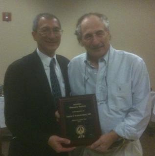
Figure 1. George Parides (left), Arizona Representative to the ATS Council of Chapter Representatives, presenting a plaque to Gerald Swartzberg (right), Arizona Thoracic Society Clinician of the Year.

A discussion was held about having a wine tasting in San Diego at the ATS International Conference. Peter Wagner (Slurping Around with PDW) has agreed to lead the conference. It was decided to extend invitations to the New Mexico, Colorado and California Thoracic Societies along with the Mayo Clinic.

A question was raised about guideline development. It was felt that we should review the Infectious Disease Society of America Valley Fever guidelines and determine if the Arizona Thoracic Society might have something to contribute.

Three cases were presented:

Lewis Wesselius from the Mayo Clinic Arizona presented a 19 year old man with shortness of breath and fever. He was seen in the Emergency Department and had a normal chest x-ray but returned 6 days later with a diffuse nodular pneumonia. Bronchoscopy with bronchoalveolar lavage revealed blood but all cultures with negative. He underwent video-assisted thorascopic lung (VATS) biopsy. Histologically the biopsy showed massive neutrophilic infiltration, hemorrhage, and small, angiocentric abscess formation. This was considered compatible with pyoderma gangrenosum of the lung (1). He had dramatic improvement with corticosteroids.

Elijah Poulos, a second year fellow at the Good Samaritan/VA program, presented a case of a non-resolving lung infiltrate in the left lower lobe after 6 weeks. CT scan showed a patchy, nodular consolidation with hazy borders. The patient was asymptomatic. Lung biopsy showed adenocarcinoma. He was referred to thoracic surgery for possible resection.  A discussion ensued reminding everyone that carcinoma is a consideration in non-resolving lung lesions and that adenocarcinoma is becoming more common (2).

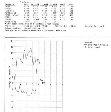
Dr. Poulos also presented a 66 year old who is retired but a semi-retired handyman/farmer who had a persistent nonproductive cough. CT scan showed a diffuse increase in interstitial markings. Pulmonary function testing revealed restrictive lung disease. Bronchoscopy with bronchoalveolar lavage was unremarkable. He was treated with fluticasone nasal spray and improved. Most advised a VATS biopsy to establish a diagnosis.

Richard A. Robbins, M.D.

References

  1. Kanoh S, Kobayashi H, Sato K, Motoyoshi K, Aida S. Tracheobronchial pulmonary disease associated with pyoderma gangrenosum. Mayo Clin Proc. 2009;84(6):555-7. [CrossRef] [PubMed] 
  2. Lortet-Tieulent J, Soerjomataram I, Ferlay J, Rutherford M, Weiderpass E, Bray F. International trends in lung cancer incidence by histological subtype: Adenocarcinoma stabilizing in men but still increasing in women. Lung Cancer. 2014 Jan 25. pii: S0169-5002(14)00044-0. [PubMed]

Reference as: Robbins RA. February 2014 Arizona Thoracic Society notes. Southwest J Pulm Crit Care. 2014;8(2):138-9. doi: http://dx.doi.org/10.13175/swjpcc026-14 PDF


Thursday
Aug292013

August 2013 Arizona Thoracic Society Notes

The August Arizona Thoracic Society meeting was held on Wednesday, 8/28/2013 at Shea Hospital beginning at 6:30 PM. There were 23 in attendance representing the pulmonary, critical care, sleep, and pathology communities.

A brief discussion was held about the audio-visual aids available. It was generally agreed that our current projector is inadequate. Judd Tillinghast will inquire about using a hospital overhead projector. If that is not possible, it was agreed to purchase a new projector.Plans for telecasting the meeting between Phoenix and Tucson continue. A trial of a link between Shea and the University in Tucson failed. Once the link is successfully established, it is hoped that the meeting can be telecasted.

There were 6 cases presented:

1. Dr. Thomas Colby, pulmonary pathologist from Mayo Clinic Arizona, presented the case of a 10 year old boy with chronic dyspnea for > 4 yrs. He had growth retardation since age 1, a skin rash since age 2 on the limbs, nail dystrophy since age 3 on hands and feet, lacrimal duct stenosis, erythematous lesions on the pinnae, phimosis, interstitial lung disease on radiography, weakly positive p-ANCA, elevated erythrocyte sedimentation rate, and hypergammaglobulinemia. He came to lung biopsy. The patient was diagnosed with dyskeratosis congenita which is a disorder of poor telomere maintenance (1). Specifically, the disease is related to one or more mutations which directly or indirectly affect the vertebrate telomerase RNA component (TERC). This patient’s manifestations are fairly typical of the disease. Short telomere length was confirmed.

2. Dr. Colby presented a second case of a 14 year old boy with a history of osteosarcoma. Pulmonary nodules developed and biopsy showed metastatic osteosarcoma. He was given systemic chemotherapy but now has residual nodules that were biopsied. One of the pulmonary nodule resembled bronchoalveolar cell carcinoma.  This is an apparent complication following chemotherapy in adolescent patients (2).

3. Dr. Colby presented the pathology of a patient from the Phoenix VA who underwent lung biopsy for interstitial disease. The pathology was typical for IgG4-related disease with a plasma cell rich lymphohistiocytic infiltrate in the bronchovascular sheath and histopathology showing diffusely stained positive for IgG4 plasma cells (3).

4. Dr. Suresh Uppalapu, a second year pulmonary fellow from Good Samaritan/VA, presented a 59 year old Sudanese male who was transferred to the Good Samaritan ICU in shock. His presenting complaints to the transferring hospital were acute mental status changes, weakness, and chills. He was intubated for hypercarbic respiratory failure. His brother related that the patient had just returned from Sudan three weeks earlier. He had a prior history of a splenectomy. He was hypothermic with a temperature of 32.3°C and a SpO2 of 91% on 100% FiO2 and PEEP of 8. His Glasgow Coma Scale was 3 (lowest possible score). He had many abnormalities on laboratory evaluation, most notably a creatinine of 5.1 mg/dL and a lactic acid of 26.3 mg/dL. The peripheral smear showed malaria parasites typical of falciparum malaria (figure 1).

Figure 1. Peripheral smear showing a gametocyte (red arrow) and trophozoites in various stages from falciparum malaria.

He developed hemoptysis and eventually expired. A preliminary autopsy report has detected aspergillosis in the lung. Invasive aspergillosis has been reported in cases of severe falciparum malaria (4).

5. Dr. Heemesh Seth, also a second year pulmonary fellow from Good Samaritan/VA, presented a case of a 57 year old man with cirrhosis secondary to hepatitis C diagnosed in 1998. He presented with a large right hydrothorax.  Multiple thoracentesis were performed (Table 1).

Table 1. Summary of multiple thoracentesis.

Blood cultures were positive for acinetobacter as was the initial culture from the thoracentesis. He was treated with cephepime. It was felt that his effusion and empyema were most likely secondary to translocation of bacteria to the pleural space from spontaneous bacterial peritonitis. A discussion ensued regarding whether to perform tube thoracostomy. Data is sparse with most literature not favoring a chest tube (5). However, in this patient’s case a chest tube was eventually inserted when he failed to improve. It drained about 2 liters of fluid but the drainage then became minimal and the tube was removed. The patient developed hepato-renal syndrome but was felt not to be a liver transplant candidate. He was transferred to hospice.

6. Dr. Seth also presented a second case of a 66 year old Hispanic man who presented with a large left pleural effusion.  He had a past medical history of systemic lupus erythematosis (SLE) with possible rheumatoid arthritis and was being treated with adalimumab, methotrexate, and prednisone. A thoracentesis was done and 2 liters of clear amber fluid was removed. Although be developed fever to 102°F he felt much better the next morning and was discharged. However, his coccidioidomycosis serologies were positive for both IgG and IgM and his complement fixation test were positive at 1:4. Pleural fluid cytology was positive for LE cells. He was continued on prednisone and treated with fluconazole. A discussion developed of whether the effusion was secondary to SLE, coccidioidomycosis, or both. No one knew data but it was felt that it was most prudent to continue the present course while following the patient and awaiting cultures.

There being no further business the meeting was adjourned at about 8:15 PM. The next meeting is scheduled for Wednesday, September 25, 6:30 PM in Phoenix at Scottsdale Shea Hospital.  

Richard A. Robbins, M.D.

References

  1. Dokal I. Dyskeratosis congenita in all its forms. Br J Haematol. 2000;110(4):768-79. [CrossRef] [PubMed] 
  2. Travis WD, Linnoila RI, Horowitz M, Becker RL Jr, Pass H, Ozols R, Gazdar A. Pulmonary nodules resembling bronchioloalveolar carcinoma in adolescent cancer patients. Mod Pathol. 1988;1(5):372-7. [PubMed]
  3. Hurley JR, Leslie KO. IgG4-Related systemic disease of the pancreas with involvement of the lung: a case report and literature review. Southwest J Pulm Crit Care. 2013;7(2):117-130. [CrossRef]
  4. Hocqueloux L, Bruneel F, Pages CL, Vachon F. Fatal invasive aspergillosis complicating severe Plasmodium falciparum malaria. Clin Infect Dis. 2000;30(6):940-2. [CrossRef] [PubMed] 
  5. Alonso JC. Pleural effusion in liver disease. Semin Respir Crit Care Med. 2010;31(6):698-705. [CrossRef] [PubMed] 

Reference as: Robbins RA. August 2013 Arizona Thoracic Society notes. Southwest J Pulm Crit Care. 2013;7(2):114-6. doi: http://dx.doi.org/10.13175/swjpcc117-13 PDF

Thursday
Jun282012

June 2012 Arizona Thoracic Society Notes

The June 2012 Arizona Thoracic Society meeting was held on 6/27/2012 at Scottsdale Shea beginning at 6:30 PM. There were 23 in attendance representing the pulmonary, critical care, sleep, pathology, infectious disease, radiology, and thoracic surgery communities.

Discussions were held regarding offering CME and partnering with other thoracic societies in the Southwest Journal of Pulmonary and Critical Care. This was endorsed by the membership. There was also discussion regarding what to discuss with Rep. David Schweikert on August 8.

Seven cases were presented:

  1. Thomas Colby, a pulmonary pathologist from the Mayo Clinic, presented a case of a 45 yo woman with a history of asthma and systemic lupus erythematosis who was found to have cysts on CT scanning. The CT scan was considered consistent with lymphangioleiomyomatosis (LAM). A lung biopsy showed only changes consistent with asthma in addition to the cysts. This case was published along with 4 similar cases by Rowan C, et al. Am J of Surg Pathol 2012;36:228–34.
  2. Jonathan Ruzi, a pulmonologist and sleep medicine specialist in Scottsdale showed an unusual flow-volume loop in an asymptomatic patient (Figure 1 below). Figure 1. Flow-volume loop of patient presented in case 2.         The cause of the obstruction was unknown but most thought this represented a type of upper airway obstruction from redundant tissue such as seen in obstructive sleep apnea or an enlarged tongue.
  3. Henry Luedy, a pulmonary fellow, presented a case of an 82 yo with cough who was a former smoker with COPD who presented with a cough. The patient presented with a consolidative process in the lingula and underwent bronchoscopy which revealed bronchial inflammation and a trace of blood in the lingula. Biopsy revealed an adenocarcinoma. Unfortunately, the pathology was not presented due to Dr. Luedy being unable to obtain the slides or images from the VA due to a clerk citing HIPAA regulations as the reason. A discussion was led by Dr. Colby on how the pathology affects the classification of these tumors as bronchoalveolar or adenocarcinoma and how there is much overlap between the classification. It was noted that educational activities are excluded from HIPAA regulations as long as the data is de-identified and there are not identifiers on a pathology slide.
  4. Tonya Whiting, a pulmonary fellow, and Manny Mathew, a pulmonologist based at Good Samaritan, presented a case of a man who developed shortness of breath while camping in the White Mountains. CT scanning revealed dense consolidation especially of the left upper lobe. He was referred for bronchoscopy but both his symptoms and consolidation resolved within 24 hours. It was felt this was a case of high altitude pulmonary edema which was somewhat unusual because high altitude pulmonary edema is unusual below 12000 feet (the patient was camping at about 9000 feet).
  5. Tonya Whiting and Allen Thomas, a pulmonologist at the VA, presented a 61 year old man with a history of polysubstance abuse and multiple lung nodules. Bronchoscopy with bronchoalveolar lavage and transbronchial biopsy was negative. Open lung biopsy revealed brochiolcentric inflammation with acute lung injury. The patient was treated with corticosteroids and the nodules resolved in 2-3 weeks. The feeling was that this represented cryptogenic organizing pneumonia (COP) presenting with multiple nodules which is a rare presentation for COP.
  6. Andrew Goldstein, a thoracic surgeon, presented a case of a 50 year old asymptomatic, nonsmoker with a huge, > 10 cm, lung tumor. The lesion was round and smooth and did not invade the chest wall on CT scan. Dr. Goldstein pointed out that pain is sensitive in predicting chest wall invasion. The tumor was resected and proved to be a carcinoid tumor.
  7. Tim Kurberski, an infectious disease specialist from Maricopa Medical Center, presented a 39 year old with a history of systemic lupus erythematosis on corticosteroids who presented with shortness and breath thought to be secondary to pulmonary edema from a cardiomyopathy. The CT scan revealed diffuse ground glass opacities. The patient also had a rash near the buttocks which was thought to be possible shingles and the chest findings possible chickenpox pneumonia. The steroids were increased but the patient failed to improve. She underwent bronchoscopy with bronchoalveolar lavage which revealed larvae consistent with Strongyloidiasis.

There being no further cases, the meeting was adjourned at 8:30 with the next meeting being a special meeting on August 8 when Rep. David Schweikert is scheduled to attend. 

Richard A. Robbins, M.D.

CCR Representative

Arizona Thoracic Society

Reference as: Robbins RA. June 2012 Arizona Thoracic Society notes. Southwest J Pulm Crit Care 2012;4:211-3. (Click here for a PDF version of the Notes)