Search Journal-type in search term and press enter
Southwest Pulmonary and Critical Care Fellowships
In Memoriam

Imaging

Last 50 Imaging Postings

(Click on title to be directed to posting, most recent listed first)

April 2024 Medical Image of the Month: Wind Instruments Player Exhibiting
   Exceptional Pulmonary Function
March 2024 Medical Image of the Month: Sputum Cytology in Patients with
   Suspected Lung Malignancy Presenting with Acute Hypoxic Respiratory
   Failure
February 2024 Medical Image of the Month: Pulmonary Alveolar Proteinosis
   in Myelodysplastic Syndrome
February 2024 Imaging Case of the Month: Connecting Some Unusual Dots
January 2024 Medical Image of the Month: Polyangiitis Overlap Syndrome
   (POS) Mimicking Fungal Pneumonia 
December 2023 Medical Image of the Month: Metastatic Pulmonary
   Calcifications in End-Stage Renal Disease 
November 2023 Medical Image of the Month: Obstructive Uropathy
   Extremis
November 2023 Imaging Case of the Month: A Crazy Association
October 2023 Medical Image of the Month: Swyer-James-MacLeod
   Syndrome
September 2023 Medical Image of the Month: Aspergillus Presenting as a
   Pulmonary Nodule in an Immunocompetent Patient
August 2023 Medical Image of the Month: Cannonball Metastases from
   Metastatic Melanoma
August 2023 Imaging Case of the Month: Chew Your Food Carefully
July 2023 Medical Image of the Month: Primary Tracheal Lymphoma
June 2023 Medical Image of the Month: Solitary Fibrous Tumor of the Pleura
May 2023 Medical Image of the Month: Methamphetamine Inhalation
   Leading to Cavitary Pneumonia and Pleural Complications
April 2023 Medical Image of the Month: Atrial Myxoma in the setting of
   Raynaud’s Phenomenon: Early Echocardiography and Management of
   Thrombotic Disease
April 2023 Imaging Case of the Month: Large Impact from a Small Lesion
March 2023 Medical Image of the Month: Spontaneous Pneumomediastinum
   as a Complication of Marijuana Smoking Due to Müller's Maneuvers
February 2023 Medical Image of the Month: Reversed Halo Sign in the
   Setting of a Neutropenic Patient with Angioinvasive Pulmonary
   Zygomycosis
January 2023 Medical Image of the Month: Abnormal Sleep Study and PFT
   with Supine Challenge Related to Idiopathic Hemidiaphragmatic Paralysis
December 2022 Medical Image of the Month: Bronchoesophageal Fistula in
   the Setting of Pulmonary Actinomycosis
November 2022 Medical Image of the Month: COVID-19 Infection
   Presenting as Spontaneous Subcapsular Hematoma of the Kidney
November 2022 Imaging Case of the Month: Out of Place in the Thorax
October 2022 Medical Image of the Month: Infected Dasatinib Induced
   Chylothorax-The First Reported Case 
September 2022 Medical Image of the Month: Epiglottic Calcification
Medical Image of the Month: An Unexpected Cause of Chronic Cough
August 2022 Imaging Case of the Month: It’s All About Location
July 2022 Medical Image of the Month: Pulmonary Nodule in the
   Setting of Pyoderma Gangrenosum (PG) 
June 2022 Medical Image of the Month: A Hard Image to Swallow
May 2022 Medical Image of the Month: Pectus Excavatum
May 2022 Imaging Case of the Month: Asymmetric Apical Opacity–
   Diagnostic Considerations
April 2022 Medical Image of the Month: COVID Pericarditis
March 2022 Medical Image of the Month: Pulmonary Nodules in the
   Setting of Diffuse Idiopathic Pulmonary NeuroEndocrine Cell Hyperplasia
   (DIPNECH) 
February 2022 Medical Image of the Month: Multifocal Micronodular
   Pneumocyte Hyperplasia in the Setting of Tuberous Sclerosis
February 2022 Imaging Case of the Month: Between A Rock and a
   Hard Place
January 2022 Medical Image of the Month: Bronchial Obstruction
   Due to Pledget in Airway Following Foregut Cyst Resection
December 2021 Medical Image of the Month: Aspirated Dental Implant
   Medical Image of the Month: Cavitating Pseudomonas
   aeruginosa Pneumonia
November 2021 Imaging Case of the Month: Let’s Not Dance
   the Twist
Medical Image of the Month: COVID-19-Associated Pulmonary
   Aspergillosis in a Post-Liver Transplant Patient
Medical Image of the Month: Stercoral Colitis
Medical Image of the Month: Bleomycin-Induced Pulmonary Fibrosis
   in a Patient with Lymphoma
August 2021 Imaging Case of the Month: Unilateral Peripheral Lung
   Opacity
Medical Image of the Month: Hepatic Abscess Secondary to Diverticulitis
   Resulting in Sepsis
Medical Image of the Month: Metastatic Spindle Cell Carcinoma of the
   Breast
Medical Image of the Month: Perforated Gangrenous Cholecystitis
May 2021 Imaging Case of the Month: A Growing Indeterminate Solitary
   Nodule
Medical Image of the Month: Severe Acute Respiratory Distress
   Syndrome and Embolic Strokes from Polymethylmethacrylate
   (PMMA) Embolization
Medical Image of the Month: Pulmonary Aspergillus Overlap Syndrome
   Presenting with ABPA, Multiple Bilateral Aspergillomas
Medical Image of the Month: Diffuse White Matter Microhemorrhages
   Secondary to SARS-CoV-2 (COVID-19) Infection
February 2021 Imaging Case of the Month: An Indeterminate Solitary
   Nodule
Medical Image of the Month: Mucinous Adenocarcinoma of the Lung
   Mimicking Pneumonia
Medical Image of the Month: Superior Vena Cava Syndrome
Medical Image of the Month: Buffalo Chest Identified at the Time of
   Lung Nodule Biopsy
November 2020 Imaging Case of the Month: Cause and Effect?
Medical Image of the Month: Severe Left Ventricular Hypertrophy
Medical Image of the Month: Glioblastoma Multiforme


For complete imaging listings click here

Those who care for patients with pulmonary, critical care or sleep disorders rely heavily on chest radiology and pathology to determine diagnoses. The Southwest Journal of Pulmonary and Critical Care publishes case-based articles with characteristic chest imaging and related pathology. The editor of this section will oversee and coordinate the publication of a core of the most important chest imaging topics. In doing so, they encourage the submission of unsolicited manuscripts. It cannot be overemphasized that both radiologic and pathologic images must be of excellent quality. As a rule, 600 DPI is sufficient for radiographic and pathologic images. Taking pictures of plain chest radiographs and CT scans with a digital camera is strongly discouraged. The figures should be cited in the text and numbered consecutively. The stain used for pathology specimens and magnification should be mentioned in the figure legend. Those who care for patients with pulmonary, critical care or sleep disorders rely heavily on chest radiology and pathology to determine diagnoses. The Southwest Journal of Pulmonary and Critical Care publishes case-based articles with characteristic chest imaging and related pathology. The editor of this section will oversee and coordinate the publication of a core of the most important chest imaging topics. In doing so, they encourage the submission of unsolicited manuscripts. It cannot be overemphasized that both radiologic and pathologic images must be of excellent quality. As a rule, 600 DPI is sufficient for radiographic and pathologic images. Taking pictures of plain chest radiographs and CT scans with a digital camera is strongly discouraged. The figures should be cited in the text and numbered consecutively. The stain used for pathology specimens and magnification should be mentioned in the figure legend.

-------------------------------------------------------------------------------------------  

Tuesday
Apr022024

April 2024 Medical Image of the Month: Wind Instruments Player Exhibiting Exceptional Pulmonary Function

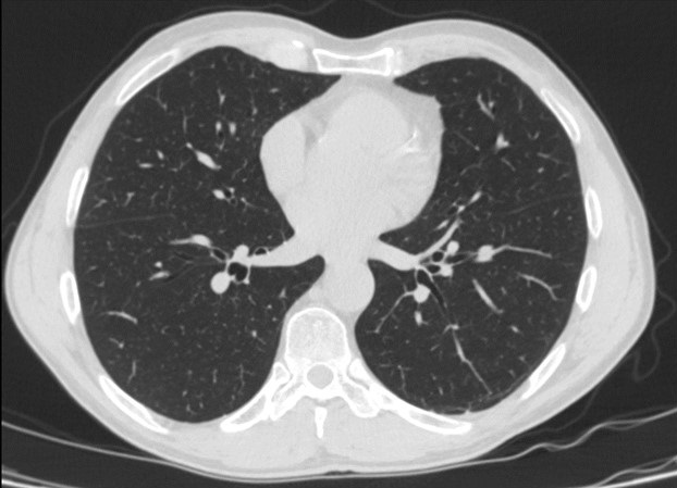
Figure 1.  Representative view from computed tomography (CT) scan (axial plane) showing clear lungs.


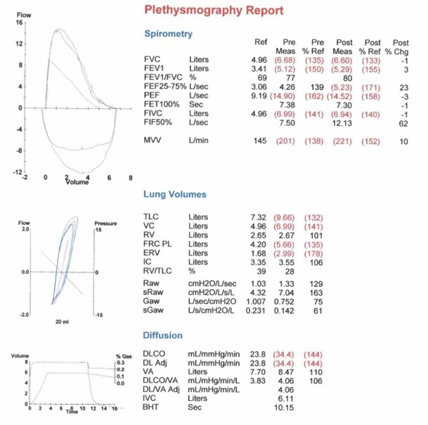
Figure 2.  Pulmonary function testing results demonstrating exceptional pulmonary function


A 64-year-old man was referred to our pulmonary clinic for evaluation of his pulmonary status.  He had a 7-year history of rheumatoid arthritis and was treated initially with steroids and subsequently maintained on methotrexate and monthly adalimumab injections. The patient reported that his rheumatoid arthritis symptoms were controlled.  He experienced no joint pain or morning stiffness at the time of evaluation. From a pulmonary perspective, he denied respiratory symptoms such as exertional shortness of breath, cough, wheezing, or chest tightness.  He reported no limitations in physical activities. The patient has an occupational history of 45-years as a welder, with exposure to dust, metal fumes, benzene, and sulfur gas. The patient also has a 15 pack-year smoking history but quit 35 years ago.

A high-resolution chest CT (Figure 1) ordered by his rheumatologist showed normal lung parenchyma. The first pulmonary function test (PFT), conducted on the initial pulmonary clinic visit, revealed lung volumes significantly higher than the reference range. This is despite the patient’s occupational history, smoking history, and the fact that he is currently on methotrexate and adalimumab therapy. The patient remained asymptomatic from a pulmonary standpoint on annual checkups. Three years later, a repeat PFT (Figure-2) demonstrated similar results.  Further history revealed that the patient had regularly used wind instruments, including the saxophone and harmonica, since high school. Initially, he played at irregular intervals, but for the last 15 years, he consistently practiced 1-2 hours daily and performed weekly at local venues.

Several studies have investigated the pulmonary effects of wind instrument playing, offering insights into the relationship between musical activities and respiratory function. Fiz et al. (1) found that maximum respiratory pressures were elevated in trumpet players.  Munn et al. (2) reported on the pulmonary function of commercial glass blowers [2]. Barbenel et al. (3) explored mouthpiece forces during trumpet playing and Kahane et al. (4) evaluated the upper airway and larynx in professional bassoon players. Cossette et al. (5) examined chest wall dynamics during flute playing. Schorr-Lesnick et al. (6) studied pulmonary function in singers and wind-instrument players [6], and Navratil et al. (7) assessed lung function in wind instrument players and glass blowers. Borgia et al. (8) provided physiological observations on French horn musicians. While existing studies present conflicting findings on the impact of wind instrument playing on respiratory function, our case adds to the growing body of evidence suggesting a potential positive correlation between long-term wind instrument training and enhanced respiratory muscle strength.

This observation prompts further exploration and investigation into the field of pulmonary rehabilitation with the hope of uncovering therapeutic benefits for individuals with chronic pulmonary conditions.

Abdulmonam Ali, MD

Pulmonary & Critical Care

SSM Health

Danville, IL USA

References

  1. Fiz JA, Aguilar J, Carreras A, Teixido A, Haro M, Rodenstein DO, Morera J. Maximum respiratory pressures in trumpet players. Chest. 1993 Oct;104(4):1203-4. [CrossRef] [PubMed]
  2. Munn NJ, Thomas SW, DeMesquita S. Pulmonary function in commercial glass blowers. Chest. 1990 Oct;98(4):871-4. [CrossRef] [PubMed]
  3. Barbenel JC, Kenny P, Davies JB. Mouthpiece forces produced while playing the trumpet. J Biomech. 1988;21(5):417-24. [CrossRef] [PubMed]
  4. Kahane JC, Beckford NS, Chorna LB, Teachey JC, McClelland DK. Videofluoroscopic and laryngoscopic evaluation of the upper airway and larynx of professional bassoon players. J Voice. 2006 Jun;20(2):297-307. [CrossRef] [PubMed]
  5. Cossette I, Monaco P, Aliverti A, Macklem PT. Chest wall dynamics and muscle recruitment during professional flute playing. Respir Physiol Neurobiol. 2008 Feb 1;160(2):187-95. [CrossRef] [PubMed]
  6. Schorr-Lesnick B, Teirstein AS, Brown LK, Miller A. Pulmonary function in singers and wind-instrument players. Chest. 1985 Aug;88(2):201-5. [CrossRef] [PubMed]
  7. Navratil M, Bejsek K. Lung function in wind instrument players and glass blowers. Ann NY Acad Sci. 1968; 155:276-83.
  8. Borgia JF, Horvath SM, Dunn FR, von Phul PV, Nizet PM. Some physiological observations on French horn musicians. J Occup Med. 1975 Nov;17(11):696-701. [PubMed]
Cite as: Ali A. April 2024 Medical Image of the Month: Wind Instruments Player Exhibiting Exceptional Pulmonary Function. Southwest J Pulm Crit Care Sleep. 2024;28(4):56-58. doi: https://doi.org/10.13175/swjpccs007-24 PDF
Saturday
Mar022024

March 2024 Medical Image of the Month: Sputum Cytology in Patients with Suspected Lung Malignancy Presenting with Acute Hypoxic Respiratory Failure

Figure 1. Axial image from a CT scan (A) showing masslike consolidation in the right lower lobe (*). Axial image from FDG-PET CT (B) showing the hypermetabolic center of the mass-like consolidation (arrow).


Figure 2. Sputum cytology low power (A) and high power (B) H&E stains showing clusters of malignant cells.


A 71-year-old woman presented with right-sided mass-like consolidation and pleural effusion on CT (Figure 1A), discovered incidentally after placement of a drug-eluting stent for coronary artery disease. The patient had a medical history significant for COPD, hypertension, hyperlipidemia, and coronary artery disease, status post a recent drug-eluting stent (less than 1 month ago). The patient received a presumptive diagnosis of pneumonia with parapneumonic effusion. Findings persisted despite multiple courses of empiric antibiotic therapy. She then underwent thoracentesis; pleural fluid was exudative; however, cytology was inconclusive. An FDG PET-CT (Figure 1B) revealed hypermetabolic activity in the right lower lobe with radiotracer activity up to 7.7 SUV concerning for malignancy. Diagnostic bronchoscopy was planned; however, her condition deteriorated suddenly the day before her planned procedure. EMS found the patient to be severely hypoxic, SpO2 in the 70s. Patient was taken by ambulance to the local emergency room.

Upon arrival, the patient was in mild respiratory distress which improved upon applying non-invasive positive-pressure ventilation. She had mild tachycardia and reduced air movement in the right lower third of the chest on physical exam. Repeat CT confirmed the persistence of the right lower lobe mass-like consolidation and moderate-sized pleural effusion. Empirical treatment for post-obstructive pneumonia was initiated. Right-sided thoracentesis again demonstrated exudative pleural fluid with negative cytology for malignancy and negative culture results. Due to concerns about her respiratory status, diagnostic bronchoscopy was abandoned. However, the patient was coughing up blood-tinged sputum which was sent for cytology (Figure 2) confirming a diagnosis of non-small cell lung cancer favoring adenocarcinoma. Immunostains performed on sections of the cell block showed malignant cells positive for CK7 and TTF1 and negative for P40 supporting the diagnosis of adenocarcinoma of lung.

This case emphasizes the importance of utilizing noninvasive testing like sputum cytology in patients with severe morbidity to help uncover underlying diagnoses. Studies and medical case reports highlighted the significance of sputum analysis in diagnosing lung cancer (1,2). Challenges posed by this case underscore the importance of considering alternative noninvasive measures to aid in making accurate diagnosis and help patient's and family understanding the underlying etiology of her persistent pneumonia )overall prognosis.

Abdulmonam Ali MD
Pulmonary & Critical Care
SSM Health
Danville, IL USA

References

  1. Thunnissen FB. Sputum examination for early detection of lung cancer. J Clin Pathol. 2003 Nov;56(11):805-10. [CrossRef] [PubMed]
  2. Ammanagi AS, Dombale VD, Miskin AT, Dandagi GL, Sangolli SS. Sputum cytology in suspected cases of carcinoma of lung (Sputum cytology a poor man's bronchoscopy!). Lung India. 2012 Jan;29(1):19-23. [CrossRef] [PubMed]
Cite as: Abdulmonam A. March 2024 Medical Image of the Month: Sputum Cytology in Patients with Suspected Lung Malignancy Presenting with Acute Hypoxic Respiratory Failure. Southwest J Pulm Crit Care Sleep. 2024;28:30-31. doi: https://doi.org/10.13175/swjpccs008-24 PDF