Search Journal-type in search term and press enter
Southwest Pulmonary and Critical Care Fellowships
In Memoriam

Imaging

Last 50 Imaging Postings

(Click on title to be directed to posting, most recent listed first)

May 2024 Medical Image of the Month: Hereditary Hemorrhagic
   Telangiectasia in a Patient on Veno-Arterial Extra-Corporeal Membrane
   Oxygenation
May 2024 Imaging Case of the Month: Nothing Is Guaranteed
April 2024 Medical Image of the Month: Wind Instruments Player Exhibiting
   Exceptional Pulmonary Function
March 2024 Medical Image of the Month: Sputum Cytology in Patients with
   Suspected Lung Malignancy Presenting with Acute Hypoxic Respiratory
   Failure
February 2024 Medical Image of the Month: Pulmonary Alveolar Proteinosis
   in Myelodysplastic Syndrome
February 2024 Imaging Case of the Month: Connecting Some Unusual Dots
January 2024 Medical Image of the Month: Polyangiitis Overlap Syndrome
   (POS) Mimicking Fungal Pneumonia 
December 2023 Medical Image of the Month: Metastatic Pulmonary
   Calcifications in End-Stage Renal Disease 
November 2023 Medical Image of the Month: Obstructive Uropathy
   Extremis
November 2023 Imaging Case of the Month: A Crazy Association
October 2023 Medical Image of the Month: Swyer-James-MacLeod
   Syndrome
September 2023 Medical Image of the Month: Aspergillus Presenting as a
   Pulmonary Nodule in an Immunocompetent Patient
August 2023 Medical Image of the Month: Cannonball Metastases from
   Metastatic Melanoma
August 2023 Imaging Case of the Month: Chew Your Food Carefully
July 2023 Medical Image of the Month: Primary Tracheal Lymphoma
June 2023 Medical Image of the Month: Solitary Fibrous Tumor of the Pleura
May 2023 Medical Image of the Month: Methamphetamine Inhalation
   Leading to Cavitary Pneumonia and Pleural Complications
April 2023 Medical Image of the Month: Atrial Myxoma in the setting of
   Raynaud’s Phenomenon: Early Echocardiography and Management of
   Thrombotic Disease
April 2023 Imaging Case of the Month: Large Impact from a Small Lesion
March 2023 Medical Image of the Month: Spontaneous Pneumomediastinum
   as a Complication of Marijuana Smoking Due to Müller's Maneuvers
February 2023 Medical Image of the Month: Reversed Halo Sign in the
   Setting of a Neutropenic Patient with Angioinvasive Pulmonary
   Zygomycosis
January 2023 Medical Image of the Month: Abnormal Sleep Study and PFT
   with Supine Challenge Related to Idiopathic Hemidiaphragmatic Paralysis
December 2022 Medical Image of the Month: Bronchoesophageal Fistula in
   the Setting of Pulmonary Actinomycosis
November 2022 Medical Image of the Month: COVID-19 Infection
   Presenting as Spontaneous Subcapsular Hematoma of the Kidney
November 2022 Imaging Case of the Month: Out of Place in the Thorax
October 2022 Medical Image of the Month: Infected Dasatinib Induced
   Chylothorax-The First Reported Case 
September 2022 Medical Image of the Month: Epiglottic Calcification
Medical Image of the Month: An Unexpected Cause of Chronic Cough
August 2022 Imaging Case of the Month: It’s All About Location
July 2022 Medical Image of the Month: Pulmonary Nodule in the
   Setting of Pyoderma Gangrenosum (PG) 
June 2022 Medical Image of the Month: A Hard Image to Swallow
May 2022 Medical Image of the Month: Pectus Excavatum
May 2022 Imaging Case of the Month: Asymmetric Apical Opacity–
   Diagnostic Considerations
April 2022 Medical Image of the Month: COVID Pericarditis
March 2022 Medical Image of the Month: Pulmonary Nodules in the
   Setting of Diffuse Idiopathic Pulmonary NeuroEndocrine Cell Hyperplasia
   (DIPNECH) 
February 2022 Medical Image of the Month: Multifocal Micronodular
   Pneumocyte Hyperplasia in the Setting of Tuberous Sclerosis
February 2022 Imaging Case of the Month: Between A Rock and a
   Hard Place
January 2022 Medical Image of the Month: Bronchial Obstruction
   Due to Pledget in Airway Following Foregut Cyst Resection
December 2021 Medical Image of the Month: Aspirated Dental Implant
Medical Image of the Month: Cavitating Pseudomonas
   aeruginosa Pneumonia
November 2021 Imaging Case of the Month: Let’s Not Dance
   the Twist
Medical Image of the Month: COVID-19-Associated Pulmonary
   Aspergillosis in a Post-Liver Transplant Patient
Medical Image of the Month: Stercoral Colitis
Medical Image of the Month: Bleomycin-Induced Pulmonary Fibrosis
   in a Patient with Lymphoma
August 2021 Imaging Case of the Month: Unilateral Peripheral Lung
   Opacity
Medical Image of the Month: Hepatic Abscess Secondary to Diverticulitis
   Resulting in Sepsis
Medical Image of the Month: Metastatic Spindle Cell Carcinoma of the
   Breast
Medical Image of the Month: Perforated Gangrenous Cholecystitis
May 2021 Imaging Case of the Month: A Growing Indeterminate Solitary
   Nodule
Medical Image of the Month: Severe Acute Respiratory Distress
Syndrome and Embolic Strokes from Polymethylmethacrylate
   (PMMA) Embolization
Medical Image of the Month: Pulmonary Aspergillus Overlap Syndrome
   Presenting with ABPA, Multiple Bilateral Aspergillomas
Medical Image of the Month: Diffuse White Matter Microhemorrhages
   Secondary to SARS-CoV-2 (COVID-19) Infection
February 2021 Imaging Case of the Month: An Indeterminate Solitary
   Nodule
Medical Image of the Month: Mucinous Adenocarcinoma of the Lung
   Mimicking Pneumonia
Medical Image of the Month: Superior Vena Cava Syndrome
Medical Image of the Month: Buffalo Chest Identified at the Time of
   Lung Nodule Biopsy
November 2020 Imaging Case of the Month: Cause and Effect?


For complete imaging listings click here

Those who care for patients with pulmonary, critical care or sleep disorders rely heavily on chest radiology and pathology to determine diagnoses. The Southwest Journal of Pulmonary and Critical Care publishes case-based articles with characteristic chest imaging and related pathology. The editor of this section will oversee and coordinate the publication of a core of the most important chest imaging topics. In doing so, they encourage the submission of unsolicited manuscripts. It cannot be overemphasized that both radiologic and pathologic images must be of excellent quality. As a rule, 600 DPI is sufficient for radiographic and pathologic images. Taking pictures of plain chest radiographs and CT scans with a digital camera is strongly discouraged. The figures should be cited in the text and numbered consecutively. The stain used for pathology specimens and magnification should be mentioned in the figure legend. Those who care for patients with pulmonary, critical care or sleep disorders rely heavily on chest radiology and pathology to determine diagnoses. The Southwest Journal of Pulmonary and Critical Care publishes case-based articles with characteristic chest imaging and related pathology. The editor of this section will oversee and coordinate the publication of a core of the most important chest imaging topics. In doing so, they encourage the submission of unsolicited manuscripts. It cannot be overemphasized that both radiologic and pathologic images must be of excellent quality. As a rule, 600 DPI is sufficient for radiographic and pathologic images. Taking pictures of plain chest radiographs and CT scans with a digital camera is strongly discouraged. The figures should be cited in the text and numbered consecutively. The stain used for pathology specimens and magnification should be mentioned in the figure legend.

-------------------------------------------------------------------------------------------  

Thursday
Nov022023

November 2023 Medical Image of the Month: Obstructive Uropathy Extremis


Figure 1. Video of CT angiography abdomen/pelvis, played caudal to cranial, obtained during assessment in the ED demonstrating obstructive uropathy with bilateral multiloculated urinomas leading to compression of the inferior vena cava. To view Figure 1  video in a separate, enlarged window click here.


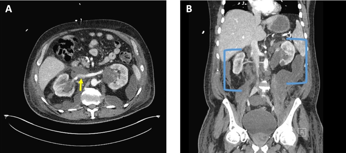
Figure 2. A: CT angiography of the abdomen, axial plane, at the level of the renal veins demonstrating compression of the inferior vena cava (yellow arrow). B: CT angiography of the abdomen and pelvis, coronal plane, demonstrating bilateral multiloculated urinomas (blue brackets). To view Figure 2 in a separate, enlarged window click here.


A 71-year-old veteran presented to the emergency department with two-weeks of progressive back pain radiating to the abdomen associated low-grade fever, nausea, and new lower extremity edema. The family reported confusion. His medical history was significant for chronic prostatitis and low-grade prostate cancer on biopsy that was lost to follow-up eleven years ago. His only reported medications were aspirin 81 mg daily and naproxen 500mg up to four times a day for his pain.

Vitals were significant for a temperature of 36.1 C, initial blood pressure of 201/74, heart rate of 128/min, respirations at 18/min with a saturation of 97% on 2L NC. Physical exam demonstrated no difference in blood pressures between arms. No abnormal heart sounds. Clear breath sounds to auscultation bilaterally. Flank tenderness to percussion and significant abdominal tenderness over the epigastric and suprapubic region with 3+ pitting edema of the bilateral lower extremities. Screening labs were notable for critical values of a WBC of 43.5 K/mL and potassium of 7 mEq/L with a creatinine of 6.5 mg/dL. He was started on esmolol and hyperkalemia temporizing therapy with the decision made to obtain an urgent contrast enhanced computed tomography of the chest, abdomen, and pelvis.

Imaging confirmed a diagnosis of severe obstructive uropathy with heterogenous prostate with nonspecific small hypodensities, marked bladder distension, hydronephroureter with ureteral wall prominence, and bilateral perinephric multiloculated fluid collections with extension into the abdominal and retroperitoneal spaces leading to indentation of the inferior vena cava (Figures 1 and 2). A Foley catheter was urgently placed with 2.5L of urine immediately relieved and prompt response in blood pressure to 130/80, and resolution of pain, altered mentation, and nausea. He was started on vancomycin and piperacillin/tazobactam for empiric coverage with three percutaneous drains subsequently placed in the multiloculated fluid collections with purulent discharge expressed. Cultures of the output demonstrated no growth. His post-obstructive diuresis was managed with replacement Lactated Ringers’ solution at 75% of the rate of Foley output. He demonstrated complete improvement in leg swelling, heart rate, and WBC and creatinine normalized to 1.1 mg/dL with discharge to home in seven days with close urology follow-up for his prostatic abnormality.

This case of obstructive uropathy extremis, probable cystocerebral syndrome (hypertension, altered mental status, and bladder distension in the elderly), and bilateral urinomas leading to inferior vena cava syndrome is unique to the literature (1). The rupture of the renal fornices, the most delicate and purported “pressure check valve” of the renal conduits, can precipitate the formation of localized urinomas within the perinephric and retroperitoneal space, most commonly unilateral from ureteral or kidney stones or tumor related obstruction, rarely bilateral from bladder outlet obstruction (2). These urinomas, as seen in this case can exert a progressively escalating pressure on the contiguous inferior vena cava, which has been described as “inferior vena cava syndrome” (3). The implications of this pressure increase are manifold, encompassing the observed clinical manifestations ranging from lower extremity edema, worsening kidney perfusion, to hemodynamic instability (3). Management centers upon decompression of the obstruction, treatment of secondary infection with source control, and compensation for post-obstructive diuresis with generally favorable prognosis if recognized early in the clinical course.

Nathan Walton MD1, Elizabeth Mata MD1, Max Hart MD1, Matthew Borchart MD2and Adnan Abbasi MBBS3

1Internal Medicine Program, University of Arizona College of Medicine – Tucson

2Department of Medicine, Pulmonary & Critical Care, University of Arizona College of Medicine – Tucson

3Department of Medicine, Pulmonary & Critical Care, Southern Arizona Veterans Affairs Health Care System (SAVAHCS) – Tucson

References

  1. Blackburn T, Dunn M. Cystocerebral syndrome. Acute urinary retention presenting as confusion in elderly patients. Arch Intern Med. 1990 Dec;150(12):2577-8. [CrossRef][PubMed]
  2. Gershman B, Kulkarni N, Sahani DV, Eisner BH. Causes of renal forniceal rupture. BJU Int. 2011 Dec;108(11):1909-11; discussion 1912. [CrossRef][PubMed]
  3. Lawrensia S, Khan YS. Inferior Vena Cava Syndrome. 2023 May 20. In: StatPearls [Internet]. Treasure Island (FL): StatPearls Publishing; 2023 Jan–. [PubMed]
Cite as: Walton N, Mata E, Hart M,  Borchart M, Abbasi A. November 2023 Medical Image of the Month: Obstructive Uropathy Extremis. Southwest J Pulm Crit Care Sleep. 2023;27(5):56-58. doi: https://doi.org/10.13175/swjpccs043-23 PDF
Wednesday
Nov012023

November 2023 Imaging Case of the Month: A Crazy Association

Parker J. Brown MD, Prasad M. Panse MD and Michael B. Gotway MD

Department of Radiology

Mayo Clinic, Arizona

Phoenix, Arizona


HPI: A 55-year-old man presents with a history of cough, poor appetite, low energy, and weight loss over the previous 6-10 months following COVID-19 infection 2 months earlier. 

PMH, SH, FH: The patient’s past medical history was positive for CVOID-19 infection 2 months earlier as well as pneumonia, not specified, in the previous year.

The patient’s past medical history was also remarkable for a 7-unit gastrointestinal hemorrhage approximately one year earlier following polypectomy for benign lesions in the transverse colon. During that hospital admission a complete blood count showed 1% blasts which prompted hematology consultation. The consulting oncologist felt the peripheral blasts were the result of a leukemoid reaction secondary to increased bone marrow stimulation owing to the patient’s acute anemia caused by the gastrointestinal hemorrhage. Macrocytosis and reticulocytosis was also noted and attributed to the same. Repeat complete blood count showed no blasts although some myelocytes, metamyelocytes, and polychromasia was noted for which follow up assessment was recommended. Serum B12 and folate levels were normal.

The patient had no prior surgeries.

The patient was not taking any prescription medications.

The patient is a non-smoker. He has no known allergies and drinks alcohol only socially and denied illicit drug use.

There was no significant family history.

Physical Examination: The patient’s physical examination showed his temperature to be 96.7°F with borderline elevated pulse rate of 95/min, a normal respiratory rate, and blood pressure of 118/67 mmHg. Room air oxygen saturation was 98%.

Initial Laboratory: A complete blood count showed a normal white blood cell count at 5.6 x10⁹/L (normal, 3.4 – 9.6 x10⁹/L), with 75% bands (normal, 50-75%). His hemoglobin and hematocrit values were 10.1 gm/dL (normal, 13.2 – 16.6 gm/dL) and 31.6% (normal, 38.3 – 48.6%). The platelet count was normal at 225 x 109/L (normal, 135 – 317 x 109/L). The patient’s serum chemistries and liver function studies were normal aside from mildly decreased total protein at 5.7 gm/dL (normal, 6.3-.9 gm/dL). The patient had an elevated anti-nuclear antibody titer at 1:320. SARS-CoV-2 PCR testing was positive.  

Radiography: Frontal chest radiography (Figure 1) was performed.

Figure 1. Frontal chest radiography at presentation.

Which of the following statements regarding this chest radiograph is accurate? (Click on the correct answer to be directed to the second of fourteen pages).

  1. Frontal chest radiography shows normal findings
  2. Frontal chest radiography shows marked cardiomegaly
  3. Frontal chest radiography shows mediastinal lymphadenopathy
  4. Frontal chest radiography shows pleural effusion
  5. Frontal chest radiography shows multifocal consolidation
Cite as: Brown PJ, Panse PM, Gotway MB. November 2023 Imaging Case of the Month: A Crazy Association. Southwest J Pulm Crit Care Sleep. 2023;27(5):47-55. doi: https://doi.org/10.13175/swjpccs046-23 PDF