Search Journal-type in search term and press enter
Southwest Pulmonary and Critical Care Fellowships
In Memoriam

Imaging

Last 50 Imaging Postings

(Click on title to be directed to posting, most recent listed first)

May 2024 Medical Image of the Month: Hereditary Hemorrhagic
   Telangiectasia in a Patient on Veno-Arterial Extra-Corporeal Membrane
   Oxygenation
May 2024 Imaging Case of the Month: Nothing Is Guaranteed
April 2024 Medical Image of the Month: Wind Instruments Player Exhibiting
   Exceptional Pulmonary Function
March 2024 Medical Image of the Month: Sputum Cytology in Patients with
   Suspected Lung Malignancy Presenting with Acute Hypoxic Respiratory
   Failure
February 2024 Medical Image of the Month: Pulmonary Alveolar Proteinosis
   in Myelodysplastic Syndrome
February 2024 Imaging Case of the Month: Connecting Some Unusual Dots
January 2024 Medical Image of the Month: Polyangiitis Overlap Syndrome
   (POS) Mimicking Fungal Pneumonia 
December 2023 Medical Image of the Month: Metastatic Pulmonary
   Calcifications in End-Stage Renal Disease 
November 2023 Medical Image of the Month: Obstructive Uropathy
   Extremis
November 2023 Imaging Case of the Month: A Crazy Association
October 2023 Medical Image of the Month: Swyer-James-MacLeod
   Syndrome
September 2023 Medical Image of the Month: Aspergillus Presenting as a
   Pulmonary Nodule in an Immunocompetent Patient
August 2023 Medical Image of the Month: Cannonball Metastases from
   Metastatic Melanoma
August 2023 Imaging Case of the Month: Chew Your Food Carefully
July 2023 Medical Image of the Month: Primary Tracheal Lymphoma
June 2023 Medical Image of the Month: Solitary Fibrous Tumor of the Pleura
May 2023 Medical Image of the Month: Methamphetamine Inhalation
   Leading to Cavitary Pneumonia and Pleural Complications
April 2023 Medical Image of the Month: Atrial Myxoma in the setting of
   Raynaud’s Phenomenon: Early Echocardiography and Management of
   Thrombotic Disease
April 2023 Imaging Case of the Month: Large Impact from a Small Lesion
March 2023 Medical Image of the Month: Spontaneous Pneumomediastinum
   as a Complication of Marijuana Smoking Due to Müller's Maneuvers
February 2023 Medical Image of the Month: Reversed Halo Sign in the
   Setting of a Neutropenic Patient with Angioinvasive Pulmonary
   Zygomycosis
January 2023 Medical Image of the Month: Abnormal Sleep Study and PFT
   with Supine Challenge Related to Idiopathic Hemidiaphragmatic Paralysis
December 2022 Medical Image of the Month: Bronchoesophageal Fistula in
   the Setting of Pulmonary Actinomycosis
November 2022 Medical Image of the Month: COVID-19 Infection
   Presenting as Spontaneous Subcapsular Hematoma of the Kidney
November 2022 Imaging Case of the Month: Out of Place in the Thorax
October 2022 Medical Image of the Month: Infected Dasatinib Induced
   Chylothorax-The First Reported Case 
September 2022 Medical Image of the Month: Epiglottic Calcification
Medical Image of the Month: An Unexpected Cause of Chronic Cough
August 2022 Imaging Case of the Month: It’s All About Location
July 2022 Medical Image of the Month: Pulmonary Nodule in the
   Setting of Pyoderma Gangrenosum (PG) 
June 2022 Medical Image of the Month: A Hard Image to Swallow
May 2022 Medical Image of the Month: Pectus Excavatum
May 2022 Imaging Case of the Month: Asymmetric Apical Opacity–
   Diagnostic Considerations
April 2022 Medical Image of the Month: COVID Pericarditis
March 2022 Medical Image of the Month: Pulmonary Nodules in the
   Setting of Diffuse Idiopathic Pulmonary NeuroEndocrine Cell Hyperplasia
   (DIPNECH) 
February 2022 Medical Image of the Month: Multifocal Micronodular
   Pneumocyte Hyperplasia in the Setting of Tuberous Sclerosis
February 2022 Imaging Case of the Month: Between A Rock and a
   Hard Place
January 2022 Medical Image of the Month: Bronchial Obstruction
   Due to Pledget in Airway Following Foregut Cyst Resection
December 2021 Medical Image of the Month: Aspirated Dental Implant
Medical Image of the Month: Cavitating Pseudomonas
   aeruginosa Pneumonia
November 2021 Imaging Case of the Month: Let’s Not Dance
   the Twist
Medical Image of the Month: COVID-19-Associated Pulmonary
   Aspergillosis in a Post-Liver Transplant Patient
Medical Image of the Month: Stercoral Colitis
Medical Image of the Month: Bleomycin-Induced Pulmonary Fibrosis
   in a Patient with Lymphoma
August 2021 Imaging Case of the Month: Unilateral Peripheral Lung
   Opacity
Medical Image of the Month: Hepatic Abscess Secondary to Diverticulitis
   Resulting in Sepsis
Medical Image of the Month: Metastatic Spindle Cell Carcinoma of the
   Breast
Medical Image of the Month: Perforated Gangrenous Cholecystitis
May 2021 Imaging Case of the Month: A Growing Indeterminate Solitary
   Nodule
Medical Image of the Month: Severe Acute Respiratory Distress
Syndrome and Embolic Strokes from Polymethylmethacrylate
   (PMMA) Embolization
Medical Image of the Month: Pulmonary Aspergillus Overlap Syndrome
   Presenting with ABPA, Multiple Bilateral Aspergillomas
Medical Image of the Month: Diffuse White Matter Microhemorrhages
   Secondary to SARS-CoV-2 (COVID-19) Infection
February 2021 Imaging Case of the Month: An Indeterminate Solitary
   Nodule
Medical Image of the Month: Mucinous Adenocarcinoma of the Lung
   Mimicking Pneumonia
Medical Image of the Month: Superior Vena Cava Syndrome
Medical Image of the Month: Buffalo Chest Identified at the Time of
   Lung Nodule Biopsy
November 2020 Imaging Case of the Month: Cause and Effect?


For complete imaging listings click here

Those who care for patients with pulmonary, critical care or sleep disorders rely heavily on chest radiology and pathology to determine diagnoses. The Southwest Journal of Pulmonary and Critical Care publishes case-based articles with characteristic chest imaging and related pathology. The editor of this section will oversee and coordinate the publication of a core of the most important chest imaging topics. In doing so, they encourage the submission of unsolicited manuscripts. It cannot be overemphasized that both radiologic and pathologic images must be of excellent quality. As a rule, 600 DPI is sufficient for radiographic and pathologic images. Taking pictures of plain chest radiographs and CT scans with a digital camera is strongly discouraged. The figures should be cited in the text and numbered consecutively. The stain used for pathology specimens and magnification should be mentioned in the figure legend. Those who care for patients with pulmonary, critical care or sleep disorders rely heavily on chest radiology and pathology to determine diagnoses. The Southwest Journal of Pulmonary and Critical Care publishes case-based articles with characteristic chest imaging and related pathology. The editor of this section will oversee and coordinate the publication of a core of the most important chest imaging topics. In doing so, they encourage the submission of unsolicited manuscripts. It cannot be overemphasized that both radiologic and pathologic images must be of excellent quality. As a rule, 600 DPI is sufficient for radiographic and pathologic images. Taking pictures of plain chest radiographs and CT scans with a digital camera is strongly discouraged. The figures should be cited in the text and numbered consecutively. The stain used for pathology specimens and magnification should be mentioned in the figure legend.

-------------------------------------------------------------------------------------------  

Friday
Feb022024

February 2024 Medical Image of the Month: Pulmonary Alveolar Proteinosis in Myelodysplastic Syndrome

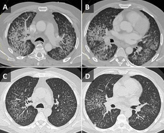
Figure 1.  Axial CT images from a contrast-enhanced chest CT performed on the patient at time of admission (A,B) demonstrate a combination of smooth septal line thickening and superimposed ground glass, resulting in a “crazy paving” appearance.  A noncontrast chest CT performed at an outside hospital 2 months earlier also demonstrates “crazy paving”; however, the findings have progressed significantly during the 2 intervening months.

A 56-year-old man presented to our institution with weight loss, fatigue, and worsening dyspnea. A chest CT (Figure 1A,B) showed a patchy, mostly central crazy-paving pattern. An outside hospital CT performed 2 months earlier was also made available for comparison (Figure 1C,D) demonstrating interval progression of these findings. The patient also had mediastinal and hilar adenopathy.  Past workup had primarily focused on the lymphadenopathy with considerations of possible lymphoma, sarcoidosis or coccidiomycosis infection. A past lymph node biopsy had revealed non-caseating granulomas and serum titers were positive for coccidiomycosis. Lymphadenopathy decreased after initiation of coccidiomycosis treatment, but symptoms and crazy paving findings continued to worsen. Further workup revealed a new diagnosis of myelodysplastic syndrome (MDS) and subsequent bronchoalveolar lavage (BAL) and histology results were consistent with secondary PAP, likely due to patient’s underlying hematologic disease.

Pulmonary alveolar proteinosis (PAP) is a rare disease characterized by the accumulation of lipoproteinaceous material in the lung alveoli. There are two types of PAP that occur in adults: Idiopathic/autoimmune and Secondary PAP. Idiopathic/autoimmune PAP is more common and is thought to result from antibody production against granulocyte-macrophage-colony-stimulating factor (GM-CSF) that regulates surfactant homeostasis. Secondary PAP results from a precipitating condition, often inhalation exposure, underlying malignancy, or immunocompromise.

The clinical manifestations of PAP are nonspecific and includes dyspnea, nonproductive cough, fatigue, and weight loss. CT may show nonspecific findings of smooth, bilateral interlobular septal thickening superimposed on a background of ground-glass opacification (crazy-paving). Diagnosis is confirmed with BAL and lung biopsy showing accumulation of eosinophilic and periodic acid-Schiff stain (PAS) positive lipoproteinaceous material within alveoli. Treatment involves pulmonary lavage for idiopathic/autoimmune PAP and treating the underlying condition for secondary PAP.

Parker Brown MD, Clint Jokerst MD, Michael Gotway MD, Matthew Stib MD

Department of Radiology

Mayo Clinic Arizona, Scottsdale, AZ

References

  1. Frazier AA, Franks TJ, Cooke EO, Mohammed TL, Pugatch RD, Galvin JR. From the archives of the AFIP: pulmonary alveolar proteinosis. Radiographics. 2008 May-Jun;28(3):883-99; quiz 915. [CrossRef][PubMed]
  2. Ioachimescu OC, Kavuru MS. Pulmonary alveolar proteinosis. Chron Respir Dis. 2006;3(3):149-59. [CrossRef] [PubMed]
  3. Presneill JJ, Nakata K, Inoue Y, Seymour JF. Pulmonary alveolar proteinosis. Clin Chest Med. 2004 Sep;25(3):593-613, viii. [CrossRef] [PubMed]
Cite as: Brown P, Jokerst C, Gotway MB, Stib M. February 2024 Medical Image of the Month: Pulmonary Alveolar Proteinosis in Myelodysplastic Syndrome. Southwest J Pulm Crit Care Sleep. 2024;28(2):18-19. doi: https://doi.org/10.13175/swjpccs004-24 PDF
Thursday
Feb012024

February 2024 Imaging Case of the Month: Connecting Some Unusual Dots

Michael B. Gotway MD

Mayo Clinic Arizona

Phoenix, AZ USA


Clinical History: A 37-year-old woman presents with abdominal pain, tongue and throat swelling, and intermittent shortness of breath and dyspnea on exertion. She also notes some pain on swallowing.

The patient’s past medical history was largely unremarkable. Her one prior surgery included cholecystectomy for cholelithiasis, and she was not taking any prescription medications.

The patient is a lifelong non-smoker, her only reported allergy due to medications containing sulfa, and she drinks alcohol only socially and denied illicit drug use.

Laboratory: A complete blood count showed a normal white blood cell count at 9.7 x 10⁹/L (normal, 3.4 – 9.6 x 10⁹/L), with an elevated absolute neutrophil count of 8.18 x 10⁹/L (normal, 1.4 – 6.6 x 10⁹/L); the percent distribution of lymphocytes, monocytes, and eosinophils was normal. Her hemoglobin and hematocrit values were 15 gm/dL (normal, 13.2 – 16.6 gm/dL) and 46% (normal, 34.9 – 44.5%). The platelet count was normal at 220 x 109/L (normal, 149 – 375 x 109/L). The patient’s serum chemistries and liver function studies were normal, including an albumin level at 4.3 gm/dL (normal, 3.5 – 5 gm/dL). SARS-CoV-2 PCR testing was negative. The erythrocyte sedimentation rate was normal at 6 mm/hr (normal, 0-29 mm/hr), although her C-reactive protein was mildly elevated at 4.8 mg/L (normal, <2 mg/L).  

Radiology: Frontal chest radiography (Figure 1) was performed.

Figure 1. Frontal chest radiography at presentation shows normal heart size, clear lungs, and no pleural abnormality.

Which of the following statements regarding this chest radiograph is accurate? (click on the correct answer to be directed to the first of twelve additional pages)

  1. Frontal chest radiography shows normal findings
  2. Frontal chest radiography shows mild cardiomegaly
  3. Frontal chest radiography shows mediastinal lymphadenopathy
  4. Frontal chest radiography shows pleural effusion
  5. Frontal chest radiography shows numerous small nodules
Cite as: Gotway MB. February 2024 Imaging Case of the Month: Connecting Some Unusual Dots. Southwest J Pulm Crit Care Sleep. 2024;28(2):9-17. doi: https:doi.org/10.13175/swjpccs001-24 PDF