Search Journal-type in search term and press enter
Southwest Pulmonary and Critical Care Fellowships
In Memoriam

Arizona Thoracic Society Notes & Videos

(Click on title to be directed to posting, most recent listed first)

January 2020 Video (Passcord TX8x3!%5)
September 2021 Video (Passcode k?6X!z@V)
June 2021 Video (Passcode S1zd7$6g)
December 2020 Arizona Thoracic Society Notes
September 2019 Arizona Thoracic Society Notes
November 2018 Arizona Thorcic Society Notes
September 2018 Arizona Thoracic Society Notes 
July 2018 Arizona Thoracic Society Notes
March 2018 Arizona Thoracic Society Notes
January 2018 Arizona Thoracic Society Notes
November 2017 Arizona Thoracic Society Notes
September 2017 Arizona Thoracic Society Notes
March 2017 Arizona Thoracic Society Notes
January 2017 Arizona Thoracic Society Notes
November 2016 Arizona Thoracic Society Notes
July 2016 Arizona Thoracic Society Notes
March 2016 Arizona Thoracic Society Notes
November 2015 Arizona Thoracic Society Notes
September 2015 Arizona Thoracic Society Notes
July 2015 Arizona Thoracic Society Notes
May 2015 Arizona Thoracic Society Notes
March 2015 Arizona Thoracic Society Notes
January 2015 Arizona Thoracic Society Notes
November 2014 Arizona Thoracic Society Notes
September 2014 Arizona Thoracic Society Notes
August 2014 Arizona Thoracic Society Notes
June 2014 Arizona Thoracic Society Notes
May 2014 Arizona Thoracic Society Notes
April 2014 Arizona Thoracic Society Notes
March 2014 Arizona Thoracic Society Notes
February 2014 Arizona Thoracic Society Notes
January 2014 Arizona Thoracic Society Notes
December 2013 Arizona Thoracic Society Notes
November 2013 Arizona Thoracic Society Notes
October 2013 Arizona Thoracic Society Notes
September 2013 Arizona Thoracic Society Notes
August 2013 Arizona Thoracic Society Notes
July 2013 Arizona Thoracic Society Notes
June 2013 Arizona Thoracic Society Notes
May 2013 Council of Chapter Representatives Notes
May 2013 Arizona Thoracic Society Notes
April 2013 Arizona Thoracic Society Notes 
March 2013 Arizona Thoracic Society Notes
March 2013 Council of Chapter Representatives Meeting 
   and “Hill Day” Notes
February 2013 Arizona Thoracic Society Notes
January 2013 Arizona Thoracic Society Notes
November 2012 Arizona Thoracic Society Notes
October 2012 Arizona Thoracic Society Notes
September 2012 Arizona Thoracic Society Notes
August 2012 Arizona Thoracic Society Notes
August 2012 Special Meeting Arizona Thoracic Society Notes
June 2012 Arizona Thoracic Society Notes
May 2012 Council of Chapter Representatives Meeting

The Arizona Thoracic Society currently has only virtual meetings about 4 times per year. These have been occurring on a Wednesday evening at 7 PM and last until about 8-8:30 PM. 

-------------------------------------------------------------------------------------

Entries in carcinoid tumor (3)

Saturday
Sep282013

September 2013 Arizona Thoracic Society Notes

The September Arizona Thoracic Society meeting was held on Wednesday, 9/25/2013 at Shea Hospital beginning at 6:30 PM. There were 13 in attendance representing the pulmonary, critical care, sleep, and pathology communities.

After a brief discussion, Gerry Swartzberg was selected as Arizona’s 2014 nominee for Clinician of the Year. 

There was 1 case presented:

Dr. Thomas Colby, pulmonary pathologist from Mayo Clinic Arizona, presented the case of a 67 year old woman with multiple pulmonary nodules. The largest was 1.2 cm CT scan. She had a fine needle aspiration of one of the nodules. The pathology revealed spindle-shaped cells which were synaptophysin + (also known as the major synaptic vesicle protein p38). Synaptophysin marks neuroendocrine tissue and on this basis the patient was diagnosed with multiple carcinoid tumors. Aguayo et al. (1) described six patients with diffuse hyperplasia and dysplasia of pulmonary neuroendocrine cells, multiple carcinoid tumorlets, and peribronchiolar fibrosis obliterating small airways. Miller and Müller (2) described a series of 25 patients that were mostly women. Eight had obliterative bronchiolitis. Many questions arose including PET positivity (variable), endobronchial spread (unknown), use of somatostatin receptor scintigraphy (unknown).

There being no further business the meeting was adjourned at about 7:30 PM. The next meeting is scheduled for Wednesday, October 23, 6:30 PM in Phoenix at Scottsdale Shea Hospital.  

Richard A. Robbins, M.D.

References

  1. Aguayo SM, Miller YE, Waldron JA Jr, Bogin RM, Sunday ME, Staton GW Jr, Beam WR, King TE Jr. Brief report: idiopathic diffuse hyperplasia of pulmonary neuroendocrine cells and airways disease. N Engl J Med. 1992;327(18):1285-8. [CrossRef] [PubMed]
  2. Miller RR, Müller NL. Neuroendocrine cell hyperplasia and obliterative bronchiolitis in patients with peripheral carcinoid tumors. Am J Surg Pathol. 1995;19(6):653-8. [CrossRef] [PubMed]

Reference as: Robbins RA. September 2013 Arizona thoracic society notes. Southwest J Pulm Crit Care. 2013;7(3):205. doi: http://dx.doi.org/10.13175/swjpcc132-13 PDF

Thursday
Jun282012

June 2012 Arizona Thoracic Society Notes

The June 2012 Arizona Thoracic Society meeting was held on 6/27/2012 at Scottsdale Shea beginning at 6:30 PM. There were 23 in attendance representing the pulmonary, critical care, sleep, pathology, infectious disease, radiology, and thoracic surgery communities.

Discussions were held regarding offering CME and partnering with other thoracic societies in the Southwest Journal of Pulmonary and Critical Care. This was endorsed by the membership. There was also discussion regarding what to discuss with Rep. David Schweikert on August 8.

Seven cases were presented:

  1. Thomas Colby, a pulmonary pathologist from the Mayo Clinic, presented a case of a 45 yo woman with a history of asthma and systemic lupus erythematosis who was found to have cysts on CT scanning. The CT scan was considered consistent with lymphangioleiomyomatosis (LAM). A lung biopsy showed only changes consistent with asthma in addition to the cysts. This case was published along with 4 similar cases by Rowan C, et al. Am J of Surg Pathol 2012;36:228–34.
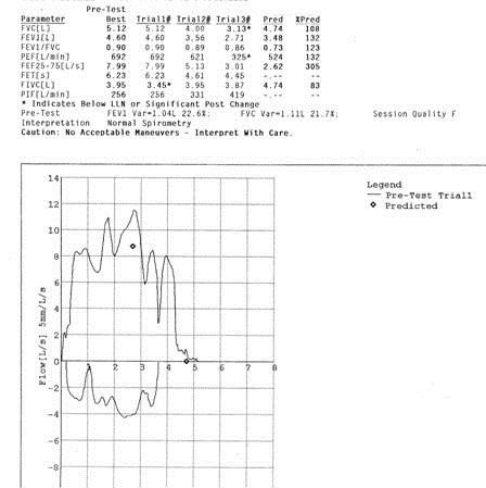
  2. Jonathan Ruzi, a pulmonologist and sleep medicine specialist in Scottsdale showed an unusual flow-volume loop in an asymptomatic patient (Figure 1 below). Figure 1. Flow-volume loop of patient presented in case 2.         The cause of the obstruction was unknown but most thought this represented a type of upper airway obstruction from redundant tissue such as seen in obstructive sleep apnea or an enlarged tongue.
  3. Henry Luedy, a pulmonary fellow, presented a case of an 82 yo with cough who was a former smoker with COPD who presented with a cough. The patient presented with a consolidative process in the lingula and underwent bronchoscopy which revealed bronchial inflammation and a trace of blood in the lingula. Biopsy revealed an adenocarcinoma. Unfortunately, the pathology was not presented due to Dr. Luedy being unable to obtain the slides or images from the VA due to a clerk citing HIPAA regulations as the reason. A discussion was led by Dr. Colby on how the pathology affects the classification of these tumors as bronchoalveolar or adenocarcinoma and how there is much overlap between the classification. It was noted that educational activities are excluded from HIPAA regulations as long as the data is de-identified and there are not identifiers on a pathology slide.
  4. Tonya Whiting, a pulmonary fellow, and Manny Mathew, a pulmonologist based at Good Samaritan, presented a case of a man who developed shortness of breath while camping in the White Mountains. CT scanning revealed dense consolidation especially of the left upper lobe. He was referred for bronchoscopy but both his symptoms and consolidation resolved within 24 hours. It was felt this was a case of high altitude pulmonary edema which was somewhat unusual because high altitude pulmonary edema is unusual below 12000 feet (the patient was camping at about 9000 feet).
  5. Tonya Whiting and Allen Thomas, a pulmonologist at the VA, presented a 61 year old man with a history of polysubstance abuse and multiple lung nodules. Bronchoscopy with bronchoalveolar lavage and transbronchial biopsy was negative. Open lung biopsy revealed brochiolcentric inflammation with acute lung injury. The patient was treated with corticosteroids and the nodules resolved in 2-3 weeks. The feeling was that this represented cryptogenic organizing pneumonia (COP) presenting with multiple nodules which is a rare presentation for COP.
  6. Andrew Goldstein, a thoracic surgeon, presented a case of a 50 year old asymptomatic, nonsmoker with a huge, > 10 cm, lung tumor. The lesion was round and smooth and did not invade the chest wall on CT scan. Dr. Goldstein pointed out that pain is sensitive in predicting chest wall invasion. The tumor was resected and proved to be a carcinoid tumor.
  7. Tim Kurberski, an infectious disease specialist from Maricopa Medical Center, presented a 39 year old with a history of systemic lupus erythematosis on corticosteroids who presented with shortness and breath thought to be secondary to pulmonary edema from a cardiomyopathy. The CT scan revealed diffuse ground glass opacities. The patient also had a rash near the buttocks which was thought to be possible shingles and the chest findings possible chickenpox pneumonia. The steroids were increased but the patient failed to improve. She underwent bronchoscopy with bronchoalveolar lavage which revealed larvae consistent with Strongyloidiasis.

There being no further cases, the meeting was adjourned at 8:30 with the next meeting being a special meeting on August 8 when Rep. David Schweikert is scheduled to attend. 

Richard A. Robbins, M.D.

CCR Representative

Arizona Thoracic Society

Reference as: Robbins RA. June 2012 Arizona Thoracic Society notes. Southwest J Pulm Crit Care 2012;4:211-3. (Click here for a PDF version of the Notes)

Thursday
Aug182011

August 2011 Arizona Thoracic Society Notes

The August Arizona Thoracic Society was held on 8/16/2011 at Scottsdale Shea beginning at 6:55 PM. There were 25 in attendance representing the pulmonary, radiology, and surgery communities.

Nine cases were presented:

1. Spontaneous Pneumothorax Secondary to Aspergilloma

Jud Tillinghast and Michael Caskey presented a case of a 65-year-old man with right upper lobe pneumonia on chest x-ray who was asymptomatic. Repeat chest x-ray showed resolution of the pneumonia, however, shortly afterwards he presented with a large right pneumothorax. CT scan of the chest showed right apical cystic changes and some areas of ground glass densities in the right upper lobe. A video-assisted thoracotomy was performed and a whitish fibrotic mass was viewed at the right apex. This was resected. Pathology revealed Aspergillus species. The patient was placed on voriconazole and made an uneventful recovery.  Drs. Tillinghast and Caskey hypothesized that one of the cystic lesions at the right apex developed an Aspergilloma and eventually ruptured causing the pneumothorax. A discussion of how long to continue the voriconazole ensued.

2. Young Woman with Hypoxemia and Hemoptysis.

Paul Conomos presented a second case of a 21-year-old woman who presented with shortness of breath, cough and hemoptysis. Her SpO2 was 87% and a CXR revealed a left lung tubular-shaped density with an enlarged left pulmonary artery. CT angiography showed several large arteriovenous (AV) malformations in the left lower lobe with several smaller lesions. The lesion was successfully embolized by coiling and the patient’s SpO2 improved to 98%.

3. Chest Masses in Identical Twins.

Dr. Conomos presented a second case of a 71-year-old woman found to have an approximate 5 cm right upper lobe mass with smaller right upper and left lower lobe nodules Biopsies of the larger right upper lobe mass and the left lower lobe nodule both revealed adenocarcinoma. Shortly thereafter, the patient’s identical twin also presented with a right middle lobe nodule- also adenocarcinoma (with bronchioloalveolar features), as well as several other suspicious-appearing pulmonary nodules, 

4. Slowly Growing Lung Mass.

Dr. Conomos presented a third case of a right lower lobe mass which was slightly enlarged compared to a previous chest x-ray in 2006. Positron emission tomography (PET) scanning showed a standardized uptake value (SUV) of 26. Needle biopsies were twice nondiagnostic. Resection revealed inflammatory  myofibroblastic tumor, also known as an inflammatory pseudotumor or plasma cell granuloma.

5. Severe Bronchiolitis Obliterans (Swyer-James Syndrome) in a 33-Year-Old.

David August presented the case of a 33-year-old man who complained of cough and had localized left upper lobe cystic bronchiectasis on chest x-ray. CT scanning also revealed left lower pulmonary artery atresia or obliteration. Discussion focused on the association of the pulmonary artery atresia / obliteration and the focal bronchiectasis.

6. Innumerable Pulmonary Cysts.

Henry Leudy and Allen Thomas presented a 63-year-old pipe smoker with a previous history of anal carcinoma who became short of breath after borrowing some bad tobacco from a friend. Chest x-ray revealed innumerable pulmonary cysts, as did thoracic CT. Images of the lung bases obtained from an abdominal CT performed in 2007 when the patient underwent resection of a 9 cm anal adenocarcinoma was unremarkable. Transbronchial biopsy showed adenocarcinoma consistent with metastatic disease. Most felt this was a very unusual radiographic appearance for metastatic disease.

7. Calcification Within a Carcinoid Tumor.

Dr. Thomas presented a second case of a 57-year-old with a tubular mass with calcification Bronchoscopy revealed a fleshy tumor in the right lower lobe bronchus which proved to be carcinoid on histological examination. Dr. Thomas presented a series that calcification was not unusual in carcinoid tumors.

8. Anti-Inflammatory Therapy for Radiation Pneumonitis.

Thomas Ardiles presented a case of a 72-year-old man who developed cough while receiving radiation therapy for mesothelioma.  His chest x-ray was compatible with radiation pneumonitis and he was begun on high dose prednisone. However, he developed mental status changes and was begun on azathioprine as the steroids were tapered without improvement. He was subsequently begun on azithromycin because of the drug’s anti-inflammatory effects with resolution of his symptoms.

9. Multiple Lung, Soft Tissue and Brain Lesions in a Patient Receiving Interferon for Hepatitis B.

Dr. Ardiles presented a second case of a 31-year-old that developed multiple bilateral small lung nodules and some scattered cutaneous and subcutaneous nodules which were noted on CT scanning. Two months later a follow up CT showed some resolution of the nodules, but most were unchanged. However, because he was complaining of headaches, brain MRI was performed and showed multiple small lesions also. Biopsy of one of the soft tissue lesions revealed cysticercosis which is due to the eggs of Taenia solium, the pork tapeworm.

The meeting adjourned at 8:30 PM.

Richard A. Robbins, MD