Search Journal-type in search term and press enter
Southwest Pulmonary and Critical Care Fellowships
In Memoriam
Social Media

Critical Care

Last 50 Critical Care Postings

(Most recent listed first. Click on title to be directed to the manuscript.)

April 2025 Critical Care Case of the Month: Being Decisive During a 
   Difficult Treatment Dilemma 
January 2025 Critical Care Case of the Month: A 35-Year-Old Admitted After
   a Fall
October 2024 Critical Care Case of the Month: Respiratory Failure in a
   Patient with Ulcerative Colitis
July 2024 Critical Care Case of the Month: Community-Acquired
   Meningitis
April 2024 Critical Care Case of the Month: A 53-year-old Man Presenting
   with Fatal Acute Intracranial Hemorrhage and Cryptogenic Disseminated
   Intravascular Coagulopathy
Delineating Gastrointestinal Dysfunction Variants in Severe Burn Injury
   Cases: A Retrospective Case Series with Literature Review
Doggonit! A Classic Case of Severe Capnocytophaga canimorsus Sepsis
January 2024 Critical Care Case of the Month: I See Tacoma
October 2023 Critical Care Case of the Month: Multi-Drug Resistant
   K. pneumoniae
May 2023 Critical Care Case of the Month: Not a Humerus Case
Essentials of Airway Management: The Best Tools and Positioning for 
   First-Attempt Intubation Success (Review)
March 2023 Critical Care Case of the Month: A Bad Egg
The Effect of Low Dose Dexamethasone on the Reduction of Hypoxaemia
   and Fat Embolism Syndrome After Long Bone Fractures
Unintended Consequence of Jesse’s Law in Arizona Critical Care Medicine
Impact of Cytomegalovirus DNAemia Below the Lower Limit of
   Quantification: Impact of Multistate Model in Lung Transplant Recipients
October 2022 Critical Care Case of the Month: A Middle-Aged Couple “Not
   Acting Right”
Point-of-Care Ultrasound and Right Ventricular Strain: Utility in the
   Diagnosis of Pulmonary Embolism
Point of Care Ultrasound Utility in the Setting of Chest Pain: A Case of
   Takotsubo Cardiomyopathy
A Case of Brugada Phenocopy in Adrenal Insufficiency-Related Pericarditis
Effect Of Exogenous Melatonin on the Incidence of Delirium and Its 
   Association with Severity of Illness in Postoperative Surgical ICU Patients
Pediculosis As a Possible Contributor to Community-Acquired MRSA
   Bacteremia and Native Mitral Valve Endocarditis
April 2022 Critical Care Case of the Month: Bullous Skin Lesions in
   the ICU
Leadership in Action: A Student-Run Designated Emphasis in
   Healthcare Leadership
MSSA Pericarditis in a Patient with Systemic Lupus
   Erythematosus Flare
January 2022 Critical Care Case of the Month: Ataque Isquémico
   Transitorio in Spanish 
Rapidly Fatal COVID-19-associated Acute Necrotizing
   Encephalopathy in a Previously Healthy 26-year-old Man 
Utility of Endobronchial Valves in a Patient with Bronchopleural Fistula in
   the Setting of COVID-19 Infection: A Case Report and Brief Review
October 2021 Critical Care Case of the Month: Unexpected Post-
   Operative Shock 
Impact of In Situ Education on Management of Cardiac Arrest after
   Cardiac Surgery
A Case and Brief Review of Bilious Ascites and Abdominal Compartment
   Syndrome from Pancreatitis-Induced Post-Roux-En-Y Gastric Remnant
   Leak
Methylene Blue Treatment of Pediatric Patients in the Cardiovascular
   Intensive Care Unit
July 2021 Critical Care Case of the Month: When a Chronic Disease
   Becomes Acute
Arizona Hospitals and Health Systems’ Statewide Collaboration Producing a 
   Triage Protocol During the COVID-19 Pandemic
Ultrasound for Critical Care Physicians: Sometimes It’s Better to Be Lucky
   than Smart
High Volume Plasma Exchange in Acute Liver Failure: A Brief Review
April 2021 Critical Care Case of the Month: Abnormal Acid-Base Balance
   in a Post-Partum Woman
First-Attempt Endotracheal Intubation Success Rate Using A Telescoping
   Steel Bougie 
January 2021 Critical Care Case of the Month: A 35-Year-Old Man Found
   Down on the Street
A Case of Athabaskan Brainstem Dysgenesis Syndrome and RSV
   Respiratory Failure
October 2020 Critical Care Case of the Month: Unexplained
   Encephalopathy Following Elective Plastic Surgery
Acute Type A Aortic Dissection in a Young Weightlifter: A Case Study with
   an In-Depth Literature Review
July 2020 Critical Care Case of the Month: Not the Pearl You Were
   Looking For...
Choosing Among Unproven Therapies for the Treatment of Life-Threatening
   COVID-19 Infection: A Clinician’s Opinion from the Bedside
April 2020 Critical Care Case of the Month: Another Emerging Cause
   for Infiltrative Lung Abnormalities
Further COVID-19 Infection Control and Management Recommendations for
   the ICU
COVID-19 Prevention and Control Recommendations for the ICU
Loperamide Abuse: A Case Report and Brief Review
Single-Use Telescopic Bougie: Case Series
Safety and Efficacy of Lung Recruitment Maneuvers in Pediatric Post-
   Operative Cardiac Patients


For complete critical care listings click here.

The Southwest Journal of Pulmonary and Critical Care publishes articles directed to those who treat patients in the ICU, CCU and SICU including chest physicians, surgeons, pediatricians, pharmacists/pharmacologists, anesthesiologists, critical care nurses, and other healthcare professionals. Manuscripts may be either basic or clinical original investigations or review articles. Potential authors of review articles are encouraged to contact the editors before submission, however, unsolicited review articles will be considered.

------------------------------------------------------------------------------------

Saturday
Oct282017

A New Interventional Bronchoscopy Technique for the Treatment of Bronchopleural Fistula

Evan Denis Schmitz, MD


Abstract

A patient receiving mechanical ventilation with multiple left hydropneumothoraces had a persistent air leak through the thoracostomy tube. The leak was temporarily resolved by interventional bronchoscopy at the bedside in the ICU. Because of the limited resources available at the hospital, a Swan-Ganz catheter was inserted into the left upper lobe bronchus, inflated and left in place. The air leak ceased and the left upper lobe bronchus was occluded with an autologous blood plug by infusing the patient’s own blood through the distal port of the catheter. The patient’s oxygenation improved significantly. The effects persisted for 2.5 hours until the air leak returned while the patient remained intubated. Such a technique may be useful when managing persistent air leaks.

Introduction

An air leak during mechanical ventilation despite the insertion of a thoracostomy tube can be detected by the bubbling of air through the air seal in the chest drainage system (1). A persistent air leak (PAL) is often defined as persistence of the air leak beyond 24 hours, which can hinder ventilation and inhibit lung expansion. Furthermore, the leak may inhibit healing of the fistula between the lung and the pleural space. Recommendations for the management of PALs include surgical repair as the gold standard for treatment (1,2). However, published anecdotal reports describe successful treatment of PALs with endobronchial insertion of fibrin sealants, ethanol injection, metal coils, Watanabe spigots and endobronchial valves. Success is also reported with chemical and autologous blood patch pleurodesis (1). We report a bedside interventional bronchoscopy technique using a Swan-Ganz catheter for the treatment of PALs while intubated and ventilated. A Swan-Ganz catheter is inserted into a lobar bronchus using direct visualization with a bronchoscope, the balloon is inflated and left in place while an autologous blood plug is created utilizing the distal port of the catheter.

Case Presentation

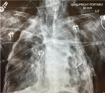
A 69-year-old man with no prior medical contact presented to the emergency department with severe shortness of breath and altered mental status. He was intubated in the emergency department for hypoxia. On arrival to the ICU he was in hypoxic respiratory failure and septic shock with a PaO2 in the 40s. His ventilator plateau pressures were 40-50 cm H2O. Chest radiography revealed moderate pneumomediastinum and multiple loculated hydropneumothoraces involving the left lung with suspected necrotic left upper lobe (Figure 1).

Figure 1. Portable AP chest x-ray showing left lung loculated hydropneumothoraces in the apex, medial and lateral walls of the left chest, subcutaneous emphysema, mediastinal emphysema and very low lung volumes. There are right apical and lower lobe areas of consolidation. A left thoracostomy tube is in place.

A 32 Fr thoracostomy tube was placed in the left intercostal space lateral to the nipple in the mid-axillary. The larger thoracostomy tube was chosen because of concern that the smaller pig-tailed catheters might not be adequate to control the leak. Plateau pressure improved to 30 cm H2O.

Despite a low tidal volume ventilator strategy and -40 cm H2O suction through the thoracostomy tube, the patient had an air leak through the thoracostomy tube which continued to bubble in the water seal chamber during both inspiration and expiration. The air leak did not improve over the ensuing 24 hours and subcutaneous emphysema worsened when attempts were made to decrease suction which was confirmed by physical exam and chest x-ray. Selective right lung ventilation led to inadequate ventilation as evidenced by increasing end-tidal CO2.

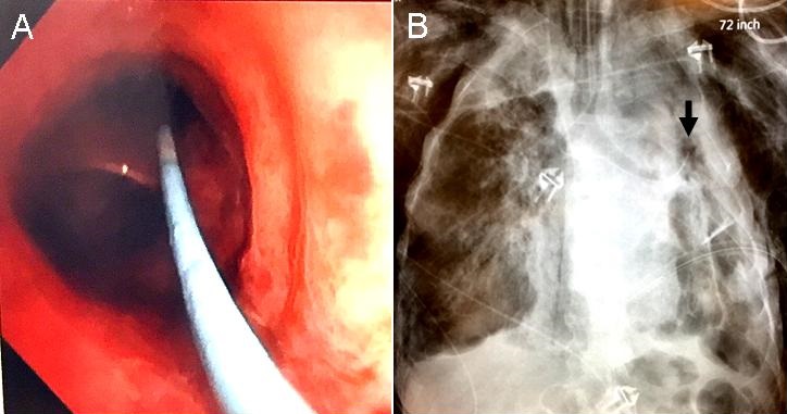
To determine and attempt to control the source of the persistent air leak, an interventional bronchoscopy was performed at bedside. Because other devices to such as metal coils, endobronchial valves, fibrin glue and a YAG laser were unavailable, a 6 Fr Swan-Ganz catheter was used. The Swan-Ganz catheter was threaded through the opening of the bronchoscope adaptor down the endotracheal tube to 3 cm above the carina. A flexible bronchoscope was then advanced along the side of the catheter through the bronchoscope adaptor and down the endotracheal tube. The catheter was not inside the working channel of the bronchoscope. The catheter was manipulated along with the bronchoscope, taking advantage of the inherent bend in the catheter, into the left mainstem bronchus and into the left upper lobe bronchus just distal to the lingular bronchus and inflated (Figure 2).

Figure 2. Panel A: Bronchoscopic view showing the Swan-Ganz catheter in the left upper lobe bronchus. Panel B: Chest x-ray confirming the Swan-Ganz catheter in the left upper lobe with the balloon inflated (arrow).

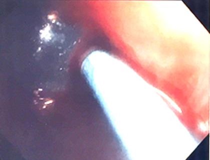
The massive air leak stopped completely. A blood plug was then created by instilling 20 ml of the patient’s own blood into the distal port of the catheter distal to the balloon along with 5 ml of 1:1000 epinephrine. The bronchoscope was used to hold the balloon in place for 10 minutes while the blood clotted. The bronchoscope was carefully removed and the catheter with the balloon inflated was left in place (Figure 3).

Figure 3. Bronchoscopic view showing the catheter passing into the left upper lobe bronchus with the surrounding blood plug.

The bronchoscope adaptor was taped post-bronchoscopy at the opening with an occlusive dressing so no air could leak around the catheter. The patient tolerated the procedure well. The air leak was successfully stopped with no evidence of worsening pneumothoraces. After PaO2 increased from the 40s on admission to the 170s after the PAL was stopped. Chest x-ray at 1 and 3 hours showed no evidence of worsening pneumothorax with the Swan-Ganz catheter still in place and inflated in the left upper lobe bronchus. After 2.5 hours, a smaller air leak did return but was present only during inspiration.

Discussion

A PAL during mechanical ventilation can be a serious complication of ventilator therapy. It can lead to poor lung expansion, ventilation/perfusion mismatch, direct extension of airway infection into the pleural space, and an inability to maintain positive end-expiratory pressure. Patients with a PAL have increased complications, including ICU readmission, pneumonia, and a longer hospital stay (3,4). Fortunately, it appears to be relatively rare. In a retrospective study only 39 out of 1,700 mechanically ventilated patients had a PAL defined as lasting for greater than 24 hours (5).

The American College of Chest Physicians guidelines published in 2001 and the 2010 British Thoracic Society guidelines on pleural disease recommend waiting for about 4 days and then seeking surgical evaluation for a PAL (2,6). It was recommended that consideration should be given to placing the thoracostomy tube to water seal rather than to suction. However, this may not be possible in patients with a large persistent air leak that complicates ventilation. In those instances, a variety of endobronchial and pleural interventions have been attempted. Although the reports are anecdotal, most achieved success with either none or minimal complications (1). There have been two basic approaches to treat PALs; sealing the air leak from the bronchial side or from the pleural side. Those therapies administered through the bronchoscope include fibrin sealant, metal coils, Watanabe spigots, synthetic hydrogel, platelet gel, endobronchial valves and YAG laser (1). Complications were infrequent and minor. Ethanolamine and ethanol have also been used but there appear to be more complications with those treatments. From the pleural side, blood patch and chemical pleurodesis have been used successfully (1). However, chemical pleurodesis might result in a trapped lung.

The technique reported here can be performed with materials available in the ICU. A torqueable guidewire can be inserted if needed to help increase the catheter stiffness and help with advancement of the catheter into the individual bronchus to identify the source of the bronchopleural fistula. Alternatives to a blood patch might include occlusion of the culprit bronchus with the patient’s own mucus and argon plasma coagulation to form a clot. A blood patch can be used to determine the potential success of a more permanent material to occlude the bronchus, such as a fibrin seal, synthetic hydrogel, laser, or before attempting endobronchial valve placement.

Conclusion

Bedside endobronchial management of PAL is feasible using a flexible bronchoscope and Swan-Ganz catheter for localization, tamponade and delivery of a blood plug.

References

  1. Dugan KC, Laxmanan B, Murgu S, Hogarth DK. Management of persistent air leaks. Chest. 2017;152(2):417-23. [CrossRef] [PubMed]
  2. Baumann MH, Strange C, Heffner JE. Management of spontaneous pneumothorax: an American College of Chest Physicians Delphi consensus statement. Chest. 2001;119(2):590-602. [CrossRef] [PubMed]
  3. Liberman M, Muzikansky A, Wright CD, et al. Incidence and risk factors of persistent air leak after major pulmonary resection and use of chemical pleurodesis. Ann Thorac Surg. 2010;89(3):891-897. [CrossRef] [PubMed]
  4. DeCamp MM, Blackstone EH, Naunheim KS, et al. Patient and surgical factors influencing air leak after lung volume reduction surgery: lessons learned from the National Emphysema Treatment Trial. Ann Thorac Surg. 2006;82(1):197-206. [CrossRef] [PubMed]
  5. Pierson DJ, Horton CA, Bates PW. Persistent bronchopleural air leak during mechanical ventilation. A review of 39 cases. Chest. 1986;90(3):321-3. [CrossRef] [PubMed]
  6. Havelock T, Teoh R, Laws D, et al. Pleural procedures and thoracic ultrasound: British Thoracic Society Pleural Disease Guideline 2010. Thorax. 2010;65(suppl 2):ii61-ii76. [CrossRef] [PubMed]

Cite as: Schmitz ED. A new interventional bronchoscopy technique for the treatment of bronchopleural fistula. Southwest J Pulm Crit Care. 2017;15(4):174-8. doi: https://doi.org/10.13175/swjpcc120-17 PDF 

Friday
Oct202017

ACE Inhibitor Related Angioedema: A Case Report and Brief Review

F. Brian Boudi, J. L. Rush, Cameron Farsar, Connie S. Chan

Carl T. Hayden VA Medical Center

University of Arizona, College of Medicine Phoenix Campus

Phoenix, AZ USA

Abstract

We present a case report of angiotensin converting enzyme (ACE) inhibitor angioedema successfully treated with icatibant (Firazyr®). The pathophysiology and treatment of ACE inhibitor angioedema is reviewed.

Introduction

Angioedema, swelling caused by a rapid increase in permeability of submucosal or subcutaneous capillaries and post-capillary venules with localized plasma extravasation, is associated with random, highly variable and often unpredictable clinical manifestations (1). Attacks are associated with significant decreased quality of life both during and between attacks, significant functional impairment and a high risk of morbidity and mortality. Angioedema can be caused by either mast cell degranulation or activation of the kallikrein-kinin cascade. ACE inhibitor-related angioedema is one the leading causes of drug-induced angioedema. While ACE inhibitor-induced angioedema is rare, awareness of this serious and potentially life-threatening complication is of great importance because of the extensive use of this class of drugs in clinical practice. Cases presenting into the emergency department because ACE inhibitors, one of the most widely prescribed medications prescribed in the United States, account for about 20-40 percent of emergency room admissions related to angioedema (1,2).

Approximately 50% of patients with ACE inhibitor-induced angioedema arise within the first week of treatment. The remainder can become symptomatic weeks, months, or even years later. The estimated incidence is likely underestimated. The actual incidence can be far higher because of poorly recognized presentation of angioedema and its sometimes-late onset. The incidence can be even higher (up to 3-fold) in certain risk groups, for instance Afro-Americans (3). It seems to have a predilection for the head, neck, lips, mouth, tongue, larynx, pharynx, and subglottal areas without urticaria (4).

Case Presentation

A 55-year-old veteran presented to the Emergency Department for the Carl T. Hayden Veterans Administration Medical Center in Phoenix Arizona with impressive angioedema. The Veteran had been taking lisinopril for 6 years and had another similar episode two months prior. The prior episode presented with facial swelling that resolved within a couple of hours. However, the present episode was accompanied by difficulty breathing and swallowing. He was begun on an allergic reaction protocol which included establishing and making sure the veteran had a patent airway, nasal trumpet, placing a peripheral intravenous catheter and starting iv fluid of sodium chloride 0.9% to keep vein open, medications of diphenhydramine 50 mg, famotidine 20 mg, methylprednisolone 125mg and 0.3 mg epinephrine subcutaneously. He was also given racemic epinephrine mixed via nebulizer and 30 mg subcutaneously of icatibant (Firazyr®), a bradykinin B2 receptor antagonist used to treat hereditary angioedema. He improved and was subsequently admitted to the intensive care unit for continued observation. The following day he was discharged with prescriptions for prednisone and orders to discontinue the use of lisinopril.

Discussion

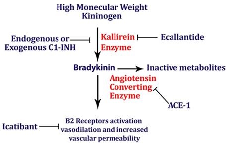
Despite newer therapies, there are no currently approved guidelines for the treatment of ACE inhibitor-induced angioedema in the United States. It is difficult to tell whether icatibant was truly effective in this case presentation as it was one of multiple therapies administered. Many causes of angioedema result from release of histamine (1). However, ACE inhibitor angioedema results from other inflammatory mediators, especially bradykinin (2) (Figure 1).

Figure 1. Simplified pathway for bradykinin-mediated angioedema showing the sites of drug activity (5).

Mast cells are not believed to be involved in this form of angioedema, and pruritus and urticaria are absent. Bradykinin-mediated angioedema, unlike histamine-mediated angioedema, frequently affects the gastrointestinal mucosa, leading to bowel wall edema and presenting with episodes of abdominal pain, nausea, vomiting, and/or diarrhea. While antihistamines and corticosteroids are often administered for treatment of angioedema, they are unlikely to have effect in ACE inhibitor induced angioedema. Epinephrine may slow (or stop) the rate of swelling. ACE inhibitor angioedema may be treated with additional drugs that act on the bradykinin pathway (e.g., icatibant, ecallantide). The recommended dose of icatibant is 30 mg administered by subcutaneous (SC) injection in the abdominal area. Additional doses may be administered in 6 hours if response is inadequate. Icatibant may decrease the time of recovery from ACE inhibitor related angioedema (6). Another ACE inhibitor should not be prescribed as the reaction is a class, not a drug specific reaction (7). Checking the complement C4 may be helpful. Patients with preexisting angioedema, including hereditary angioedema caused by C1 esterase inhibitor deficiency, are predisposed to develop angioedema in response to ACE inhibitors (8).

ACE inhibitor induced angioedema remains a disorder without a clear treatment modality for reduction of symptoms. The primary therapeutic interventions remain removal of the offending agent and airway management when indicated. The use of icatibant may be effective in the management of ACE inhibitor related angioedema; however, its efficacy and benefits have not been clear in the small studies published thus far. There have been three randomized trials evaluating the use of icatibant in ACE inhibitor angioedema. Interestingly, the first study found icatibant to be effective while the more recent and larger studies found no significant difference in time to recovery (3, 6, 9-12). Icatibant is costly with a wholesale price of $9,000-$11,000 and may not be available at all hospitals. Given its questionable outcomes data, icatibant may not appropriate in all medical centers. This is especially important since off-label use may not be covered by insurers. 

References

  1. Stone C Jr, Brown NJ. Angiotensin-converting enzyme inhibitor and other drug-associated angioedema. Immunol Allergy Clin North Am. 2017 Aug;37(3):483-495. [CrossRef] [PubMed]
  2. Guyer AC, Banerji A. ACE inhibitor-induced angioedema. UpToDate. June 27, 2017. Available at: https://www.uptodate.com/contents/an-overview-of-angioedema-clinical-features-diagnosis-and-management#H30 (requires subscription, accessed 9/18/17).
  3. Straka BT, Ramirez CE, Byrd JB, et al. Effect of bradykinin receptor antagonism on ACE inhibitor-associated angioedema. J Allergy Clin Immunol. 2017;140:242-248.e2. [CrossRef] [PubMed]
  4. Sabroe R, Black A. Angiotensin-converting enzyme (ACE) inhibitors and angio-oedema. Br J Dermatol. 1997;1:153–8. [CrossRef] [PubMed]
  5. Shenvi C, Serrano K. New treatments for angioedema. Emergency Physicians Monthly. 9/12/16. Available at: http://epmonthly.com/article/new-treatments-angioedema/ (accessed 10/20/17).
  6. Baş M, Greve J, Stelter K, et al. A randomized trial of icatibant in ACE-inhibitor-induced angioedema. N Engl J Med. 2015 Jan 29;372(5):418-25. [CrossRef] [PubMed]
  7. Johnsen SP, Jacobsen J, Monster TB, Friis S, McLaughlin JK, Sørensen HT.Risk of first-time hospitalization for angioedema among users of ACE inhibitors and angiotensin receptor antagonists. Am J Med. 2005;1:1428-9. [CrossRef] [PubMed]
  8. Orfan N, Patterson R, Dykewicz M. Severe angioedema related to ACE inhibitors in patients with a history of idiopathic angioedema. JAMA. 1990;1:1287-9. [CrossRef] [PubMed]
  9. Sinert R, Levy P, Bernstein JA, et al.Randomized trial of icatibant for angiotensin-converting enzyme inhibitor-induced upper airway angioedema. J Allergy Clin Immunol Pract. 2017 Sep-Oct;5(5):1402-9.e3. [CrossRef] [PubMed]
  10. Culley CM, DiBridge JN, Wilson GL Jr. Off-label use of agents for management of serious or life-threatening angiotensin converting enzyme inhibitor-induced angioedema. Ann Pharmacother. 2016 Jan;50(1):47-59 [CrossRef] [PubMed]
  11. Fok JS, Katelaris CH, Brown AF, Smith WB. Icatibant in angiotensin-converting enzyme (ACE) inhibitor-associated angioedema. Intern Med J. 2015 Aug;45(8):821-7. [CrossRef] [PubMed]
  12. Riha HM, Summers BB, Rivera JV, Van Berkel MA. Novel therapies for angiotensin-converting enzyme inhibitor-induced angioedema: a systematic review of current evidence. J Emerg Med. 2017 Sep 19. pii: S0736-4679(17)30489-4. [CrossRef] [PubMed]

Cite as: Boudi FB, Rush JL, Farsar C, Chan CS. ACE inhibitor related angioedema: a case report and brief review. Southwest J Pulm Crit Care. 2017;15(4):165-8. doi: https://doi.org/10.13175/swjpcc114-17 PDF 

Thursday
Oct052017

Tumor Lysis Syndrome from a Solitary Nonseminomatous Germ Cell Tumor

Brandon T. Nokes, MD1

Rodrigo Cartin-Ceba, MD2

Joseph Farmer, MD2

Alyssa B. Chapital, MD, PhD2


1Hospital Internal Medicine and 2Division of Critical Care

Mayo Clinic Arizona

Phoenix, AZ USA


Abstract

Spontaneous tumor lysis syndrome is a rare clinical entity, which typically occurs in the context of rapidly proliferating hematologic malignancies. Tumor lysis syndrome in solid organ malignancies is even rarer, and typically provoked by cytotoxic treatment regimens. We describe a case of spontaneous tumor lysis of a solitary metastatic brain lesion from a nonseminomatous germ cell tumor. This case is unique in that spontaneous tumor lysis from a brain metastasis of a solid organ malignancy has never been reported, and spontaneous tumor lysis in a nonseminomatous germ cell tumor is exceedingly rare.

Case Report

A 31-year-old gentleman was admitted to our facility after developing status epilepticus and consequently, being involved in a MVA. Imaging revealed a 3.5cm right frontal brain lesion with surrounding edema, but no other acute intracranial pathology. The patient was intubated, sedated, and transferred to critical care for further treatment. His past medical history was notable for primary surgical resection of a T1N0M0 nonseminomatous germ cell tumor in March 2015, followed by detection of a 2.5cm lung nodule in September 2015, with concurrent beta-human chorionic gonadotropin (HCG) and alpha-fetoprotein (AFP) biochemical recurrence. He underwent 4 cycles of bleomycin, etoposide, and cisplatin (BEP).

A head CT revealed a 4cm x 3.5cm right frontal lesion with surrounding edema (Figure 1).

Figure 1. T2 Axial MRI showing 4 cm x 3.5 cm lesion with associated vasogenic edema.

Dexamethasone 4mg every 6 hours was initiated for treatment of vasogenic edema. Laboratory studies were significant for a white blood cell count elevated at 19.3 x10⁹/L, international normalized ratio (INR) 1.34, partial thromboplastin time (PTT) 26.2 seconds, and prothrombin time (PT) 16.1 seconds. Plasma lactate was elevated at 30.6mmol/L. Bicarbonate was 6mmol/L with an anion gap of 45, glucose 186mg/dL, BUN 15.2mg/dL, and creatinine was 2.0mg/dL. Urine drug screen was negative. His AFP was 7.4ng/mL and beta-HCG was 13IU/L. Over the following 24 hours, the patient experienced decreased urine output. A bedside ultrasound reveals normal IVC collapse. Further lab assessment revealed a CK within normal limits and a urinalysis showed the presence of 11 to 20 RBCs, 4 to 10 WBCs and some granular casts as well as trace protein. His phosphorus was 8.9, calcium 8.1, and uric acid was 13mg/dL. His lactate dehydrogenase levels were also elevated at 271 U/L.

Due to concern of tumor lysis syndrome, the patient was initiated on rasburicase, which was followed by maintenance allopurinol 300mg daily. However, due to worsening renal failure, the patient was started on hemodialysis. He was taken to the operating room the following morning for immediate surgical resection of his brain metastasis; no evidence of residual disease was seen on follow-up imaging (Figure 2).

Figure 2. T2 Axial MRI status post a right frontal craniotomy and gross total resection of the previously noted mass. Small amount of blood noted within the resection cavity. Residual vasogenic edema persists in the white matter surrounding the operative bed.

Repeat chest, abdomen and pelvis imaging did not show any additional metastatic lesions.

In the following days, he was subsequently extubated, transferred to the floor, and continued hemodialysis, eventually fully recovering his renal function. Ultimately, he was discharged with outpatient follow-up for additional chemotherapy planning after physical rehabilitation.

Discussion

Tumor lysis syndrome (TLS) can be subdivided into laboratory TLS and clinical TLS, as defined by the Cairo-Bishop diagnostic criteria (1). Spontaneous TLS can occur in solid organ malignancies (1). TLS in solid organ malignancies is provoked by chemotherapy or radiation therapy, which creates massive cell lysis and elaboration of intracellular potassium, phosphate, and uric acid as well as hypocalcemia, which can lead to renal failure and cardiac dysrhythmias (1). LDH is also elevated. TLS can also be thought of as being provoked, either by ongoing chemotherapy or a decrease in effective circulating volume, or unprovoked. It is rare for TLS to occur in nonseminomatous germ cell tumors. Only 2 case reports have been published regarding spontaneous TLS in nonseminomatous germ cell tumors (2,3). Our case is most likely a spontaneous TLS. To date, no reports have been published regarding spontaneous TLS from a solitary brain metastasis from a nonseminomatous germ cell tumor. Further, no cases have been reported regarding tumor lysis from a solitary brain metastasis of any solid organ malignancy.

The occurrence of TLS in solid organ malignancies is thought to occur secondary to rapid cellular proliferation that exceeds the available blood supply for a tumor, leading to tumor ischemia and diffuse tumor cell necrosis. The biochemical milieu elaborated from these necrotic cells can result in end-organ pathology.

The treatment of TLS is contingent upon the rate of cancer progression and whether there is evidence of end-organ damage. Importantly and ideally, patients can be stratified into intermediate, moderate, or high-risk of developing TLS based on their malignancy type and rate of cancer progression, such that TLS may be prevented with prophylactic hydration, electrolyte monitoring and allopurinol or rasburicase (4,5). Biochemical TLS alone can be treated with IV hydration and allopurinol, a xanthine oxidase inhibitor which potentially halts TLS progression. When there is end-organ damage, rasburicase (a recombinant urate oxidase) is the first-line treatment along with aggressive hydration (5). Additional therapies are directed towards minimizing sequelae of TLS (i.e. calcium gluconate for hyperkalemia associated EKG changes or emergent dialysis for acute renal failure). There is no role for urinary alkalinization.

We were fortunate in that our patient had a great outcome, owing to early detection and aggressive intervention, and we implore our fellow physicians to be mindful of TLS as a possible clinical outcome in all malignancies, irrespective of its clinical rarity.

References

  1. Mirrakhimov AE, Ali AM, Khan M, Barbaryan A. Tumor lysis syndrome in solid tumors: an up to date review of the literature. Rare Tumors. 2014;6(2):5389. [CrossRef] [PubMed]
  2. D'Alessandro V, Greco A, Clemente C, et al. Severe spontaneous acute tumor lysis syndrome and hypoglycemia in patient with germ cell tumor. Tumori. 2010;96(6):1040-3. [PubMed]
  3. Pentheroudakis G, O'Neill VJ, Vasey P, Kaye SB. Spontaneous acute tumour lysis syndrome in patients with metastatic germ cell tumours. Report of two cases. Support Care Cancer. 2001;9(7):554-7. [CrossRef] [PubMed]
  4. Feres GA, Salluh JI, Ferreira CG, Soares M. Severe acute tumor lysis syndrome in patients with germ-cell tumors. Indian J Urol. 2008;24(4):555-7. [CrossRef] [PubMed]
  5. Coiffier B, Altman A, Pui CH, Younes A, Cairo MS. Guidelines for the management of pediatric and adult tumor lysis syndrome: an evidence-based review. J Clin Oncol. 2008;26(16): 2767-78. [CrossRef] [PubMed]

Cite as: Nokes BT, Cartin-Ceba R, Farmer J, Chapital AB. Tumor lysis syndrome from a solitary nonseminomatous germ cell tumor. Southwest J Pulm Crit Care. 2017;15(4):148-50. doi: https://doi.org/10.13175/swjpcc107-17 PDF

Monday
Oct022017

October 2017 Critical Care Case of the Month

Margaret Ragland, MD1

Carolyn H. Welsh, MD1,2


Pulmonary Sciences and Critical Care Medicine

1University of Colorado Anschutz Medical Campus and 2VA Eastern Colorado Health Care System

Denver, Colorado USA


History of Present Illness

A 42-year-old man with a history of intravenous heroin abuse and chronic hepatitis C infection presents to the emergency department (ED) with recurrent abdominal pain. The pain was dull, epigastric, and did not radiate. The pain worsened after eating, but the timing after eating that it worsened was inconsistent. He had nausea but no vomiting. His bowel movements were normal without constipation, diarrhea, or melena.   

He had presented to another ED multiple times with this same pain over the past six weeks. He does not know what the work-ups revealed, but was discharged from the emergency department each time. He received supportive care including fluids and analgesics, but the pain would always recur a few hours after returning home.

He went to a third ED a few weeks ago with bilateral testicular pain after which he was discharged home with acetaminophen for pain.

Past Medical History, Family History, and Social History

His past medical history is notable for bipolar disorder. He takes no prescribed medications and does not know his family’s medical history. He is a current every day smoker, has no history of heavy alcohol use, and uses intravenous heroin but no other recreational drugs.

Current Medications

Acetaminophen a few times a day for abdominal pain.

Review of Systems

He notes subjective fevers, poor appetite, and an 8 pound unintentional weight loss over the past six weeks.

Physical Exam

Vital signs are notable for hypertension to 158/91 mm Hg. Other vitals are within normal limits.

On exam, he is an ill appearing middle aged man who appears very uncomfortable. His abdomen is nondistended. He has normal bowel sounds and epigastric tenderness with a tender, smooth liver edge palpable just under the costal margin. He has decreased sensation to light touch in his toes with no skin changes. Toes are warm with capillary refill less than two seconds.

Laboratory Evaluation

CBC reveals a leukocytosis to 23,600 cells/mcL with 80% neutrophils; eosinophils are normal. Hemoglobin and platelet counts are normal. Sodium is 128 mmol/L with a bicarbonate of 30 mmol/L and creatinine of 0.64 mmol/L. AST 155 U/L, ALT 137 U/L, with a total bilirubin 1.1 mmol/L. Albumin is 1.8 g/L. INR is 1.9. Urinalysis showed 1+ protein.

What additional laboratory evaluation is indicated at this time? (Click on the correct answer to proceed to the second of six pages)

  1. Acetaminophen level
  2. Hepatitis B viral (HBV) serologies
  3. Lipase
  4. 1 and 3
  5. All of the above

Cite as: Ragland M, Welsh CH. October 2017 critical care case of the month. Southwest J Pulm Crit Care. 2017;15(4):131-7. doi: https://doi.org/10.13175/swjpcc113-17 PDF 

Saturday
Sep022017

September 2017 Critical Care Case of the Month

James T. Dean III, MD

Tyler R. Shackelford, DO

Michel Boivin, MD

Division of Pulmonary, Critical Care and Sleep Medicine

University of New Mexico School of Medicine

Albuquerque, NM USA


Critical Care Case of the Month CME Information

Members of the Arizona, New Mexico, Colorado and California Thoracic Societies and the Mayo Clinic are able to receive 0.25 AMA PRA Category 1 Credits™ for each case they complete. Completion of an evaluation form is required to receive credit and a link is provided on the last panel of the activity. 

0.25 AMA PRA Category 1 Credit(s)™

Estimated time to complete this activity: 0.25 hours 

Lead Author(s): James T. Dean III, MD.  All Faculty, CME Planning Committee Members, and the CME Office Reviewers have disclosed that they do not have any relevant financial relationships with commercial interests that would constitute a conflict of interest concerning this CME activity.

Learning Objectives: As a result of completing this activity, participants will be better able to:

  1. Interpret and identify clinical practices supported by the highest quality available evidence.
  2. Establish the optimal evaluation leading to a correct diagnosis for patients with pulmonary, critical care and sleep disorders.
  3. Translate the most current clinical information into the delivery of high quality care for patients.
  4. Integrate new treatment options for patients with pulmonary, critical care and sleep related disorders.

Learning Format: Case-based, interactive online course, including mandatory assessment questions (number of questions varies by case). Please also read the Technical Requirements.

CME Sponsor: University of Arizona College of Medicine

Current Approval Period: January 1, 2017-December 31, 2018

Financial Support Received: None


A 73-year-old man presented with a three-day history of diffuse abdominal pain, decreased urine output, nausea and vomiting. His past medical history included diabetes, coronary artery disease, hypertension and chronic back pain. The patient reported being started on hydrochlorothiazide, furosemide, pregabalin and diclofenac within the last week in addition to his long-standing metformin prescription.

Initial vitals were significant for tachypnea, tachycardia to 120 bpm, hypothermia to 35ºC and hypotension with a blood pressure of 70/40 mm Hg. Physical exam was remarkable for bilateral lung wheezing and significant respiratory distress. Laboratory examination was concerning for a pH of 6.85, pCO2 of < 5mmHg, serum lactate of 27mmol/l, WBC of 15.6 x10⁶ cells/cc and a serum creatinine of 8.36 mg/dl. A chest X-ray showed evidence of mild pulmonary edema and a CT of the abdomen did not show any acute pathology.

What is the most likely etiology of the patient’s severe acidosis? (Click on the correct answer to proceed to the second of four pages)

  1. Diabetic ketoacidosis
  2. Ethylene glycol poisoning
  3. Metformin-associated lactic acidosis
  4. Septic shock

Cite as: Dean JT III, Shackelford TR, Boivin M. September 2017 critical care case of the month. Southwest J Pulm Crit Care. 2017;15(3):100-3. doi: https://doi.org/10.13175/swjpcc101-17 PDF