Search Journal-type in search term and press enter
Southwest Pulmonary and Critical Care Fellowships
In Memoriam
Social Media

Critical Care

Last 50 Critical Care Postings

(Most recent listed first. Click on title to be directed to the manuscript.)

April 2025 Critical Care Case of the Month: Being Decisive During a 
   Difficult Treatment Dilemma 
January 2025 Critical Care Case of the Month: A 35-Year-Old Admitted After
   a Fall
October 2024 Critical Care Case of the Month: Respiratory Failure in a
   Patient with Ulcerative Colitis
July 2024 Critical Care Case of the Month: Community-Acquired
   Meningitis
April 2024 Critical Care Case of the Month: A 53-year-old Man Presenting
   with Fatal Acute Intracranial Hemorrhage and Cryptogenic Disseminated
   Intravascular Coagulopathy
Delineating Gastrointestinal Dysfunction Variants in Severe Burn Injury
   Cases: A Retrospective Case Series with Literature Review
Doggonit! A Classic Case of Severe Capnocytophaga canimorsus Sepsis
January 2024 Critical Care Case of the Month: I See Tacoma
October 2023 Critical Care Case of the Month: Multi-Drug Resistant
   K. pneumoniae
May 2023 Critical Care Case of the Month: Not a Humerus Case
Essentials of Airway Management: The Best Tools and Positioning for 
   First-Attempt Intubation Success (Review)
March 2023 Critical Care Case of the Month: A Bad Egg
The Effect of Low Dose Dexamethasone on the Reduction of Hypoxaemia
   and Fat Embolism Syndrome After Long Bone Fractures
Unintended Consequence of Jesse’s Law in Arizona Critical Care Medicine
Impact of Cytomegalovirus DNAemia Below the Lower Limit of
   Quantification: Impact of Multistate Model in Lung Transplant Recipients
October 2022 Critical Care Case of the Month: A Middle-Aged Couple “Not
   Acting Right”
Point-of-Care Ultrasound and Right Ventricular Strain: Utility in the
   Diagnosis of Pulmonary Embolism
Point of Care Ultrasound Utility in the Setting of Chest Pain: A Case of
   Takotsubo Cardiomyopathy
A Case of Brugada Phenocopy in Adrenal Insufficiency-Related Pericarditis
Effect Of Exogenous Melatonin on the Incidence of Delirium and Its 
   Association with Severity of Illness in Postoperative Surgical ICU Patients
Pediculosis As a Possible Contributor to Community-Acquired MRSA
   Bacteremia and Native Mitral Valve Endocarditis
April 2022 Critical Care Case of the Month: Bullous Skin Lesions in
   the ICU
Leadership in Action: A Student-Run Designated Emphasis in
   Healthcare Leadership
MSSA Pericarditis in a Patient with Systemic Lupus
   Erythematosus Flare
January 2022 Critical Care Case of the Month: Ataque Isquémico
   Transitorio in Spanish 
Rapidly Fatal COVID-19-associated Acute Necrotizing
   Encephalopathy in a Previously Healthy 26-year-old Man 
Utility of Endobronchial Valves in a Patient with Bronchopleural Fistula in
   the Setting of COVID-19 Infection: A Case Report and Brief Review
October 2021 Critical Care Case of the Month: Unexpected Post-
   Operative Shock 
Impact of In Situ Education on Management of Cardiac Arrest after
   Cardiac Surgery
A Case and Brief Review of Bilious Ascites and Abdominal Compartment
   Syndrome from Pancreatitis-Induced Post-Roux-En-Y Gastric Remnant
   Leak
Methylene Blue Treatment of Pediatric Patients in the Cardiovascular
   Intensive Care Unit
July 2021 Critical Care Case of the Month: When a Chronic Disease
   Becomes Acute
Arizona Hospitals and Health Systems’ Statewide Collaboration Producing a 
   Triage Protocol During the COVID-19 Pandemic
Ultrasound for Critical Care Physicians: Sometimes It’s Better to Be Lucky
   than Smart
High Volume Plasma Exchange in Acute Liver Failure: A Brief Review
April 2021 Critical Care Case of the Month: Abnormal Acid-Base Balance
   in a Post-Partum Woman
First-Attempt Endotracheal Intubation Success Rate Using A Telescoping
   Steel Bougie 
January 2021 Critical Care Case of the Month: A 35-Year-Old Man Found
   Down on the Street
A Case of Athabaskan Brainstem Dysgenesis Syndrome and RSV
   Respiratory Failure
October 2020 Critical Care Case of the Month: Unexplained
   Encephalopathy Following Elective Plastic Surgery
Acute Type A Aortic Dissection in a Young Weightlifter: A Case Study with
   an In-Depth Literature Review
July 2020 Critical Care Case of the Month: Not the Pearl You Were
   Looking For...
Choosing Among Unproven Therapies for the Treatment of Life-Threatening
   COVID-19 Infection: A Clinician’s Opinion from the Bedside
April 2020 Critical Care Case of the Month: Another Emerging Cause
   for Infiltrative Lung Abnormalities
Further COVID-19 Infection Control and Management Recommendations for
   the ICU
COVID-19 Prevention and Control Recommendations for the ICU
Loperamide Abuse: A Case Report and Brief Review
Single-Use Telescopic Bougie: Case Series
Safety and Efficacy of Lung Recruitment Maneuvers in Pediatric Post-
   Operative Cardiac Patients


For complete critical care listings click here.

The Southwest Journal of Pulmonary and Critical Care publishes articles directed to those who treat patients in the ICU, CCU and SICU including chest physicians, surgeons, pediatricians, pharmacists/pharmacologists, anesthesiologists, critical care nurses, and other healthcare professionals. Manuscripts may be either basic or clinical original investigations or review articles. Potential authors of review articles are encouraged to contact the editors before submission, however, unsolicited review articles will be considered.

------------------------------------------------------------------------------------

Monday
Feb022015

February 2015 Critical Care Case of the Month: A Bloody Mess

Mily Sheth, MD

Carmen Luraschi, MD

Matthew P. Schreiber, MD, MHS


University of Nevada School of Medicine: Las Vegas

Department of Internal Medicine

Division of Pulmonary/Critical Care

Las Vegas, NV


History of Presenting Illness:

A 23-year-old Ethiopian woman with a known history of systemic lupus erythematosus (SLE) but of unknown duration presented with the chief complains of cough and generalised weakness for 1 week. She had a recent history of travelling to Ethiopia 3 months ago for 3 weeks. She complained of subjective fevers and one episode of blood tinged sputum. She also complained of fatigue and an episode of syncope which prompted her hospitalization.

PMH, SH and FH:

The patient has a past medical history of SLE diagnosed in Ethiopia of which no records were available. She is a student and denied alcohol, smoking or drug abuse. She denied any family history of autoimmune disorders. She did not take any medications at home.

Physical Examination:

Initial admission vital signs were temperature of 100.5 F, heart rate of 130, respiratory rate of 30 and blood pressure of 92/48. Oxygen saturation was 96% on 2 L/min via nasal cannula.

She appeared to be in moderate distress but was speaking in full sentences. Skin examination revealed a malar rash on her face. Her upper and lower extremities had excoriated plaques. Her anterior chest had flat non blanchable, macular rash. CVS examination revealed tachycardia without any murmurs. Respiratory exam was positive for bilaterally diffuse bronchial breath sounds. The remainder of her exam was within normal limits.

Laboratory and Radiology:

CBC: WBC 6.7 million cells/mcL, hemoglobin 7.1 g/dL, hematocrit 20.9, platelet 160,000 cells/mcL

Renal panel: within normal limits.

Troponin 0.01, creatine kinase 457 U/L, lactic acid 1.1 mm/L, HIV non-reactive

Liver function tests: AST 288 U/L, ALT 93 U/L alkaline phosphatase 136 IU/L, total bilirubin 0.9 mg/dL

Radiography:

Her initial chest x-ray is shown in figure 1. It was interpreted as showing diffuse pulmonary infiltrates, right lung greater than left. No pleural effusions. No pneumothorax.

Figure 1. Initial chest x-ray.

In a patient with these characteristics, which other test(s) would you order? (Click on the correct answer to proceed to the second of five panels)

  1. Arterial blood gases and lactic acid
  2. Cardiac angiogram
  3. Computed tomography (CT) of the chest without contrast
  4. VATS lung biopsy
  5. All of the above

Reference as: Sheth M, Luraschi C, Schreiber MP. February 2015 critical care case of the month: a blood mess. Southwest J Pulm Crit Care. 2015;10(2):63-9. doi: http://dx.doi.org/10.13175/swjpcc148-14 PDF

Tuesday
Jan062015

Physical Examination in the Intensive Care Unit: Opinions of Physicians at Three Teaching Hospitals

Rodrigo Vazquez, MD1

Cristina Vazquez Guillamet, MD1

Mohamed Adeel Rishi, MD2

Jorge Florindez, MD4

Priya S Dhawan, MD3

Sarah E. Allen, MD1

Constantine A Manthous, MD5

Geoffrey Lighthall MD, PhD6

Affiliations: University of New Mexico School of Medicine1 Albuquerque NM. McNeal Hospital2, Berwyn Il. Mayo Clinic Arizona3, Scottsdale AZ. Bridgeport Hospital4, Bridgeport CT. Yale School of Medicine5, New Haven CT. Stanford University School of Medicine6, Stanford CA.

Study Sites: Stanford University Medical Center, Stanford CA. McNeal Hospital, Berwyn Il and Bridgeport Hospital, Bridgeport CT.


Abstract

Purpose: Technological advances in intensive care unit may lead physicians to question or omit portions of the physical exam. Our goal is to assess the opinions of intensive care unit physicians about physical examination in modern day medicine.

Methods: Subjects included physicians on medical intensive care unit teams at one university hospital and two university-affiliated teaching hospitals. Participants responded to an interview divided into two sections: (1) A semi-structured interview including open-ended questions on the management of four critical care scenarios and on the utility of physical exam; (2) Multiple-choice questions about physical exam.

Main Results: The response rate was 100%. A total of 122 individuals, 16(13%) attendings, 24(20%) fellows and 82(67%) residents, responded. Half 61 (50%) considered physical examination to be of limited utility in the intensive care unit. Fifteen percent of answers to the clinical scenarios were reasoned based on physical examination. Most extended the definition of physical examination to include data derived from monitoring 119(97%), life support 121(99%) and bedside imaging devices 112(92%). Residents 45(37%), students 35(29%) and nurses 35(29%) were recognized as the team members who examine patients the most.

Conclusion: Physical examination was considered useful by half of the physicians. Percussion is the least appreciated component. The role of nurses examining patients is recognized. A new definition of physical examination that extends beyond the patient to include monitoring, life support and bedside imaging is proposed to revitalize bedside clinical medicine.

For accompanying editorial click here.

Introduction

Physical examination is one of the mainstays of clinical activities at the bedside. The many maneuvers and signs of the physical exam were developed and described over the last two centuries when most patients presented in advanced states of disease with obvious physical examination findings (1). In the last few decades, advances in fields of imaging, laboratory and bedside monitoring technologies have increased expectations for early and accurate diagnoses, often before physical exam findings become apparent (2).

In Critical Care Units (ICU) patients have severe presentations of diseases, making it likely to encounter diagnostic physical examination findings; however ICUs have easy access to imaging and automated physiologic measurements that may lead physicians to question, or omit portions, of the physical examination.

In this context, we sought the opinions of physicians working in intensive care units about physical examination in modern day medicine.

Material and Methods

The study was divided into two sections. The first is based on mixed methods analysis (qualitative and quantitative) of semi-structured interviews with open-ended questions. The second is based on the quantitative analysis of multiple-choice questions.

Setting

The study was conducted in 2011, in three ICUs in three states.  One of three hospitals (Stanford) was a 32-bed closed medical-surgical unit in a university medical center hospital. The other two hospitals were 16-17 bed closed medical units at university-affiliated community teaching hospitals. All three hospitals had postgraduate residencies in Internal Medicine and Pulmonary and Critical Care Medicine, and were equipped with electronic medical records systems and computer acquisition and storage of bedside data. At the time of the study, Stanford had begun an initiative to increase the use and appreciation of physical examination in its medical school (3). No other confounding variables were apparent. The Investigational Review Boards of each center approved the protocol independently and waived the need for written consent.

Eligible subjects included residents, critical care fellows, and attending physicians on ICU rotations. Investigators approached all candidates for possible participation; subjects were informed that the study would evaluate their approach to diagnosis and treatment of ICU patients but not about its specific focus on physical examination.

Data collection

Demographic data collected included age, gender, type of specialty and subspecialty training, and level of training.

First section

Semi-structured interviews on how four hypothetical ICU clinical vignettes would be managed; questions were chosen by consensus of the authors and responses were open-ended.

Subjects were presented with this introductory statement: “I’m going to present you with four clinical scenarios. I would like you to explain how you would manage these clinical scenarios in real life. This is not an exam. We are just interested in how physicians practice.” The following four case scenarios were then introduced: “You have to manage an Intensive Care Unit patient with: 1) hypoxemia, 2) hypotension, 3) dyspnea and 4) oliguria. What would you do?”

After discussing management of the case scenarios, subjects were then asked: “What’s your opinion about the utility of physical exam in the ICU?”

Second section

Multiple-choice questions were then asked of each subject:

- How frequently do you examine your patients? Answer choices (or options): “Always, sometimes, never”

-Who do you think examines their patients the most? Answer choices: “Attendings, fellows, residents, students or nurses”

 -Which data obtained at the bedside should be included in an updated definition of physical exam in the ICU? Inspection? palpation? percussion? auscultation? venous lines? arterial line data? ventilator data?, bedside ultrasonography? Answer options: “Strongly agree, agree, disagree, strongly disagree”

The interview was pilot tested in six subjects to verify subjects had a clear understanding of the questions. Investigators performing the interviews were the same within each center and were trained prior to subject enrollment. Investigators read the questions in the same order. Subsequent questions were not revealed until the previous question had been answered. Responses were recorded and transcribed.

Analysis

Data analysis used a mixed-methods approach for the first section. The transcriptions were analyzed looking for key actions or ideas around which the rest of the response was organized, we called these “codes”. For example if an individual answered: “I would auscultate the lungs, order an arterial blood gas and a chest radiograph ” the codes would be lung auscultation, arterial blood gas and chest radiograph. After analyzing all the answers we decided to group “codes” into categories and report them as percentages of all the answers provided, four clinical scenarios per each of the 122 participants. For the previous example the categories would include: auscultation, laboratory test and radiology.

Mention of the physical exam was categorized as: a) mentions physical examination (e.g. “ I would examine the patient…”), b) mentions physical examination or the intention to go to the bedside, c) describes a reasoned physical examination (e.g.“ I would auscultate the lungs, if I heard wheezes then I would…”).

Illustrative comments were highlighted. The findings of each interviewer were checked against each other. In case of discrepancy, answers were compared to reach a consensus.

The analysis of the second section was quantitative.

We used the statistical package Stata 11. Chi2 was used to compare rates. Factorial logistic regression and logistic regression were used to evaluate categorical and continuous data when appropriate. A p -value of <0.05 was considered significant.

Results

A total of 122 individuals were approached for study participation and all agreed to participate. The subjects included 16 (13%) attendings, 24 (20%) fellows and 82 (67%) residents. The average age was 32 years (range 24-65). There were 79 (65%) males. Most respondents had Internal Medicine training 116 (95%) and had attended medical school in the US 76 (62%).

First section

Clinical scenarios

Categories identified during the responses to the clinical scenarios are summarized in Table 1.

We report mention of the physical exam in three not mutually exclusive categories: a) mentions physical examination (e.g. “ I would examine the patient…”), b) mentions physical examination or the intention to go to the bedside, c) describes a reasoned physical examination (e.g.“ I would auscultate the lungs, if I heard wheezes then I would…”).

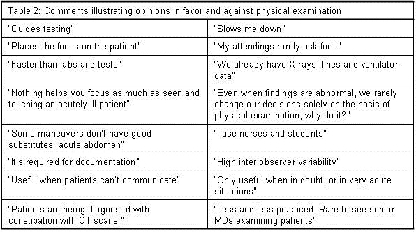
Answers to “What’s your opinion about the utility of physical exam in the ICU?” The physical exam was considered to be of “limited utility” by 61(50%) of the respondents. Table 2 includes answers that illustrate opinions for and against the utility of the physical exam.

According to the respondents, the components of the physical exam that remain useful in the intensive care unit are: a) general appearance; b) the neurological exam; c) abdominal exam since there are no adequate monitoring devices; d) anterior auscultation of the chest to detect pneumothoraces, effusions or cardiac murmurs; and e) examination of the skin.

Second section, multiple-choice questions

A. How frequently do you examine your patients?

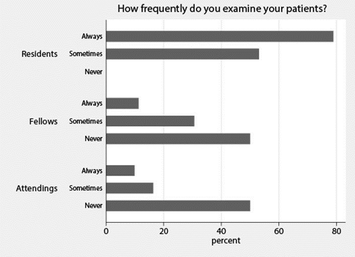
Figure 1. Histogram with the percentages of each of the possible answers to question: How frequently do you examine your patients?

B. Who do you think examines patients the most? (Figure 2).

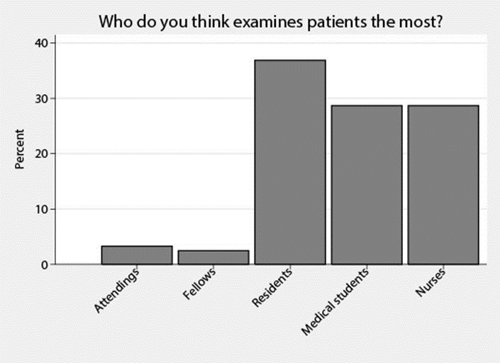
Figure 2. Histogram with the percentages for each of the possible answers to question: Who do you think examines patients the most?

C. Which data obtained at the bedside should be included in an updated definition of physical exam in the ICU?

At least 90% of the respondents agreed to include data obtained through inspection 112 (100%), auscultation 118 (97%), data from ventilators 121 (99 %), arterial lines 120 (98%), central lines 118 (97%), and bedside ultrasound 112(92%). Palpation 107(88%) and percussion 79 (65%) did not exceed the 90% threshold.

Besides the intergroup comparisons noted above, there were no statistically significant differences between responses based on level of training, age, gender, location of medical school training or hospital (p>0.05).

Discussion

We report the opinion of 122 physicians with regard to physical examination in the intensive care unit and the way they reported using the physical exam in four hypothetical clinical scenarios. We found that half of the physicians reported they considered physical exam useful, but only 15 percent mentioned physical exam in deducing answers to the clinical scenarios. Percussion was the least appreciated component of the physical exam. There was generalized agreement that the inclusion of data derived from bedside imaging, monitoring, and life support devices into an updated definition of physical examination would be valuable. Nurses and students were recognized as the team members who examined their patients the most. Study participants provided explanations behind their opinions.

The findings that only 50% of the physicians found the standard physical examination to be useful, and that only 15 % mentioned the physical exam in deducing their case scenario answers suggests a low appreciation for the standard physical exam. Available literature on the utility of the physical exam supports our current study findings. In one study the time spent at the bedside during clinical rounds was down to 11% from an historical 75%, with most of the time spent in hallways or conference rooms (4,5). In an ethnographic study, residents felt it was unnecessary to examine their patients in the ICU as long as they had monitoring and a good nurse (6).

Perceptions from patients and the general public support our findings that use of the physical exam is low. In a questionnaire to ambulatory patients, 56 patients perceived 113 omissions in their physician visit, the most common omissions being those related to what they felt were missed portions of the physical examination (7). Mass media has produced the following headlines: Physician revives a dying art: the physical; Is physical exam facing extinction? and Not on the Doctors’ checklist but touch matters (8-10).

Comments by our study participants help explain these findings. Participants had more confidence in the accuracy of data provided by monitoring devices and imaging than in findings of their physical exams.  Some participants mentioned that it was difficult to convince peers to change management based on physical examination findings alone. Participants also reported there was lack of role modeling physicians performing physical exams.

Attending and fellow physicians were perceived as the team members who examine their patients the least; Attending and fellows validated this perception in reporting examining their patients sometimes or never in more than half of the cases.

An alternative explanation for this perception about the senior team members could be related to differences in diagnostic reasoning between trainees and more experienced physicians (11). Students and residents probably relay more in hypothetic deductive reasoning and collect larger amounts of data, including a more detailed physical examination, to reach a diagnosis. As they become more experienced and start working as fellows and attendings the use of short cuts, heuristics, increases and they reach a diagnosis with smaller amounts of data. Time constraints also make them relay in the information relayed by more junior team members.

Whatever the reason for this perception about senior team members may be, it explains, at least in part, the atrophy of physical examination skills during residency training (12), and feeds a downward spiral with graduates that examine their patients less and less.

Residents, nurses and students on the other hand were recognized as the ones who examined their patients the most. This observation expands the role of nurses in the ICU, and if confirmed by others could change the allocation of responsibilities in the multidisciplinary ICU team.

Although until now our discussion portrays the current poor standings of physical examination in the ICU, we also found hope.

Despite the small proportion of participants basing their reasoning on physical examination during the clinical scenarios, most responses included going to the bedside and almost all participants agreed on extending the physical examination beyond the patient to include data derived from monitoring, life support and bedside imaging devices.

Just as Laennec revolutionized bedside diagnosis with the introduction of the stethoscope (13), a new standardized definition of physical examination including these new bedside diagnostic tools, has the potential to greatly enrich clinical medicine and would reflect the practice of modern medicine better (14,15). Critical care medicine is in a privilege position to lead this conceptual change and spread it to other specialties.

Our study has several limitations. First of all, it describes opinions and behaviors in theoretical scenarios and the answers provided may not correlate with true physician practices. The order and choice of the clinical scenarios and questions may have biased the respondents negatively against the physical exam. In support of our results, once the participants learned about the focus of the study one would have expected an attempt to offer better impressions of themselves with an “over reporting” bias towards the physical examination. However, our results pointed in the opposite direction. It does not correlate the use of physical examination to outcomes.

In regards to the composition of the respondents, residents conformed the great majority. Although one could argue that this limits the generalizability of the results, the proportion of residents, fellows and attending physicians mimics that found in the ICU teams at the participating institutions. Our findings may not be generalized to hospitals in areas with limited resources.

Finally its main limitation and at the same time its main virtue is the generation of new questions that will require new studies with direct observation of team practices and their correlation to patient outcomes.

Conclusion

Physical examination was considered useful by half of the physicians. Percussion is the least appreciated component. The role of nurses examining patients is recognized. A new definition of physical examination that extends to include monitoring, life support and bedside imaging is proposed to revitalize bedside clinical medicine.

Conflict of interests: The authors declare that they have no conflict of interest.

References

  1. Walker HK, Hall WD, Hurst JW, Walker HK. The origins of the history and physical examination 3rd ed. Boston, MA: Butterworths; 1990.
  2. Berenguer J, Bruguera M, Gervas J, et al. The Physician of the Future. El Metge del Futur. Fundacion Educaion Medica, Barcelona, Spain; 2009. Available at: http://www.econ.upf.edu/~ortun/publicacions/MedicoDelFuturo.pdf (accessed 1/6/15).
  3. Verghese A, Horwitz RI. In praise of the physical examination. BMJ. 2009;339:b5448. [CrossRef] [PubMed]
  4. LaCombe MA. On bedside teaching. Ann Intern Med. 1997;126(3):217-20. [CrossRef] [PubMed]
  5. Miller M, Johnson B, Greene HL, Baier M, Nowlin S. An observational study of attending rounds. J Gen Intern Med. 1992;7(6):646-8. [CrossRef] [PubMed]
  6. Bosk CL. Forgive and remember: managing medical failure. 2nd ed. Chicago, IL. University of Chicago Press; 2003. [CrossRef]
  7. Kravitz RL, Callahan EJ. Patients' perceptions of omitted examinations and tests: A qualitative analysis. J Gen Intern Med. 2000;15(1):38-45. [CrossRef] [PubMed]
  8. Knox R. The Fading Art Of The Physical Exam. National Public Radio. 2010. Available at: http://www.npr.org/player/v2/mediaPlayer.html?action=1&t=1&islist=false&id=129931999&m=129984296 (accessed 1/6/15).
  9. Ofri D. Not on the doctor's checklist but touch matters. The New York Times. August 2, 2010.
  10. Grady D. Physician revives a dying art: the physical. The New York Times. October 11, 2010. Available at: http://www.nytimes.com/2010/10/12/health/12profile.html?pagewanted=all&_r=0 (accessed 1/6/15).
  11. Sackett DL, Tugwell P, Guyatt GH. Clinical epidemiology: a basic science for clinical medicine, 2nd ed. Boston: Little, Brown; 1991.
  12. Mangione S, Nieman LZ. Cardiac auscultatory skills of internal medicine and family practice trainees. A comparison of diagnostic proficiency. JAMA. 1997;278(9):717-22. [CrossRef] [PubMed]
  13. Laennec R. Traite de l’auscultation mediate. Bull Acad Natl Med. 1819;151:393-398.
  14. COBATRICE Domain 2: diagnosis: assessment, investigation, monitoring and data interpretation. Available at: http://www.cobatrice.org/Data/ModuleGestionDeContenu/PagesGenerees/en/02-competencies/0B-diagnosis/8.asp (accessed 1/6/15).
  15. American Association of Chest Physicians. Critical care ultrasonography. http://www.chestnet.org/Education/Advanced-Clinical-Training/Certificate-of-Completion-Program/Critical-Care-Ultrasonography (accessed 1/6/15).

Reference as: Vazquez R, Vazquez Guillamet C, Adeel Rishi M, Florindez J, Dhawan PS, Allen SE, Manthous CA, Lighthall G.  Physical examination in the intensive care unit: opinions of physicians at three teaching hospitals. Southwest J Pulm Crit Care. 2015;10(1):34-43. doi: http://dx.doi.org/10.13175/swjpcc165-14 PDF

Saturday
Jan032015

Analysis of a Fatal Left Ventricular Assist Device Infection: A Case Report and Discussion

Neal Stuart Gerstein, MD FASE1

Henry G. Chou, MD2

Andrew Lewis Dixon, MD1


1Department of Anesthesiology & Critical Care Medicine

University of New Mexico

Albuquerque, NM

2Department of Anesthesiology

Cedars-Sinai Medical Center

Los Angeles, CA


Introduction

Left ventricular assist device (VAD) therapy is an increasingly utilized treatment as a bridge to heart transplantation or as long-term destination therapy. Recent reports show there is a 22% - 32% incidence of VAD-associated infections with staphylococci and nosocomial gram-negative bacilli being the most common causative organisms (1,2). These organisms are often found in intensive care units, where they have the highest proportion of resistance, thus exposing already critically ill patients to the possibility of resistant organism VAD-associated infections (3). Mortality rates exceed 60% when sepsis develops in a patient with a continuous flow left VAD and infection is the number one cause of death in those awaiting cardiac transplantation (4,5). With continued left VAD use clinicians will likely see multidrug-resistant (MDR) or even pandrug-resistant organism VAD-associated infections. Clinicians need to be prepared to manage such an intimidating entity.

Case Report

We report a case of a 25 year-old male with a pandrug-resistant Pseudomonas aeruginosa VAD-associated infection. The patient’s medical history is significant for a diagnosis of idiopathic dilated cardiomyopathy refractory to maximal medical therapy requiring implantation of a HeartMate II (Thoratec Co., Pleasanton, CA, USA) continuous flow left VAD (Figure 1).

Figure 1. HeartMate II® left VAD schematic (reprinted with the permission of Thoratec Co., Pleasanton, CA, USA).

His course was complicated with multiple hospital admissions for recurrent VAD-associated infections and numerous episodes of P. aeruginosa bacteremia that had been treated with a multitude of antipseudomonal antibiotics. He presented to our hospital for management of severe volume overload in the setting of VAD-associated infections. Transesophageal echocardiography demonstrated a left ventricular ejection fraction of 24% with severe left and right ventricular dilatation. Chest x-ray revealed cardiomegaly and multiple devices including the left VAD (Figure 2).

Figure 2. Chest X-ray demonstrating an enlarged cardiac silhouette, the HeartMate II axial pump (*) with inflow (down arrow, ↓) and outflow (up arrow, ↑) cannulas, biventricular pacer with leads in right atrium (A), coronary sinus (B), and right ventricle (C) (dashed arrows).

Blood cultures revealed MDR P. aeruginosa; except for showing intermediate sensitivity to tobramycin there was resistance to all antimicrobials tested. In vitro synergy testing revealed modest bacterial inhibition when only colistin, fosfomycin, imipenem, and tobramycin were combined. After maximizing medical therapy, multiple left VAD pocket washings and implantation of tobramycin beads followed. Intraoperative findings included an encapsulated infection around the driveline and obvious infection of the left VAD pocket. Repeat blood cultures showed P. aeruginosa had developed resistance to all antimicrobials including tobramycin. Subsequently the left VAD was explanted and the patient was transitioned to an extracorporeal membrane oxygenator (ECMO) in attempt to clear the infection. He was then transitioned to a TandemHeart (CardiacAssist Inc., Pittsburgh, PA, USA), a percutaneous LVAD, as he was not dependent on ECMO for oxygenation. He was able to clear the bacteremia after removal of the infected HeartMate II while on colistin, fosfomycin, tobramycin, azithromycin and rifampin, but was not able to clear the remaining left VAD pocket infection, which again spread systemically. Despite maximal medical and surgical interventions, he died from profound septic shock and multisystem organ failure. To date this is the first known case of a pandrug-resistant P. aeruginosa VAD-associated infection reported in the literature.

Discussion

P. aeruginosa organisms have intrinsic resistance to numerous broad spectrum antibiotics, and can easily develop acquired resistance to most if not all available antimicrobial agents (3). Risk factors for the development of pandrug-resistant P. aeruginosa include previous treatment with antipseudomonal antibiotics and prolonged treatment times. Given our patient had multiple P. aeruginosa infections, treated with multiple rounds of antipseudomonal antibiotics, it is not surprising that pandrug-resistance developed. Few therapeutic options are available for treatment and no new agents are available to evade the known resistance mechanisms. Treatment can be optimized using synergistic combination therapy, which may be the only medical management option in patients with pandrug-resistant P. aeruginosa infections. Some have suggested that rifampin in combination with colistin may be a promising approach (3). Some experts recommend in vitro synergy testing when an organism is resistant to currently recommended antibiotic regimens (6,7). However, a recent review of antibiotic therapy for gram-negative infections describes the utility of in vitro synergy testing equivocal in the context of Pseudomonas infection (8). We managed our patient with combination therapy; however, not until pandrug-resistant P. aeruginosa was isolated did we introduce rifampin in combination with colistin.

A recent review of VAD-associated infections showed the majority were managed without surgical intervention; only 13% required surgical debridement and only in cases of severe infection and/or failed conservative treatment was left VAD explantation required. Since this case there has been a proposed algorithm for management of VAD-associated infections (2); our management, though prior to published guidelines, was in step with the algorithm. Of note, there was no discussion of explanting an left VAD to ECMO to aid in clearing a resistant infection. We felt this was a rational option given our inability to clear the infection. It is unclear as to exactly why our patient was never able to fully clear his infection. Given the patient’s other pre-existing extensive cardiac hardware (i.e. implanted pacer), it is possible that he remained colonized even after maximal surgical and medical therapy. Though speculative, it is possible that removing all foreign material may have allowed for complete infection clearance.

Aside from aggressive medical and surgical management, systolic heart failure with VAD-associated infections may be effectively managed with heart transplantation (9). Our consensus was that this option was neither in the best interest of the patient nor the best use of available resources given the severity of his condition.

Conclusion

Clinicians will continue to see VAD-associated infections with resistant organisms. To minimize adverse outcomes, including VAD-associated infection, prudent patient selection and timing of VAD placement is paramount, as VAD’s placed in critically ill patients have been consistently associated with adverse outcomes (10).

References

  1. Gordon RJ, Weinberg AD, Pagani FD, Slaughter MS, Pappas PS, Naka Y, Goldstein DJ, Dembitsky WP, Giacalone JC, Ferrante J, Ascheim DD, Moskowitz AJ, Rose EA, Gelijns AC, Lowy FD. Prospective, multicenter study of ventricular assist device infections. Circulation. 2013;127:691-702. [CrossRef] [PubMed]
  2. Nienaber JJ, Kusne S, Riaz T, Walker RC, Baddour LM, Wright AJ, Park SJ, Vikram HR, Keating MR, Arabia FA, Lahr BD, Sohail MR. Clinical manifestations and management of left ventricular assist device-associated infections. Clin Infect Dis. 2013;57:1438-48. [CrossRef] [PubMed]
  3. Zavascki AP, Carvalhaes CG, Picao RC, Gales AC. Multidrug-resistant Pseudomonas aeruginosa and Acinetobacter baumannii: resistance mechanisms and implications for therapy. Expert Rev Anti Infect Ther. 2010;8:71-93. [CrossRef] [PubMed]
  4. Topkara VK, Kondareddy S, Malik F, Wang IW, Mann DL, Ewald GA, Moazami N. Infectious complications in patients with left ventricular assist device: etiology and outcomes in the continuous-flow era. Ann Thorac Surg. 2010;90:1270-7. [CrossRef] [PubMed]
  5. Bartoli CR, Demarest CT, Khalpey Z, Takayama H, Naka Y. Current management of left ventricular assist device erosion. J Card Surg. 2013;28:776-82. [CrossRef] [PubMed]
  6. Balaji V, Jeremiah SS, Baliga PR. Polymyxins: Antimicrobial susceptibility concerns and therapeutic options. Indian J Med Microbiol. 2011;29:230-42. [CrossRef] [PubMed]
  7. Martis N, Leroy S, Blanc V. Colistin in multi-drug resistant Pseudomonas aeruginosa blood-stream infections: a narrative review for the clinician. J Infect. 2014;69:1-12. [CrossRef] [PubMed]
  8. Tamma PD, Cosgrove SE, Maragakis LL. Combination therapy for treatment of infections with gram-negative bacteria. Clin Microbiol Rev. 2012;25:450-70. [CrossRef] [PubMed]
  9. Prendergast TW, Todd BA, Beyer AJ, 3rd, Furukawa S, Eisen HJ, Addonizio VP, Browne BJ, Jeevanandam V. Management of left ventricular assist device infection with heart transplantation. Ann Thorac Surg. 1997;64:142-7. [CrossRef] [PubMed]
  10. Lietz K, Long JW, Kfoury AG, Slaughter MS, Silver MA, Milano CA, Rogers JG, Naka Y, Mancini D, Miller LW. Outcomes of left ventricular assist device implantation as destination therapy in the post-REMATCH era: implications for patient selection. Circulation. 2007;116:497-505. [CrossRef] [PubMed] 

Reference as: Gerstein NS, Chou HG, Dixon AL. Analysis of a fatal left ventricular assist device infection: a case report and discussion. Southwest J Pulm Crit Care. 2015;10:16-20. doi: http://dx.doi.org/10.13175/swjpcc139-14 PDF 

Friday
Jan022015

January 2015 Critical Care Case of the Month: Who's Your Momma?

Carlos Hartmann, MD

Layth Al-Jashaami, MD

Timothy T. Kuberski, MD


Department of Medicine

Maricopa Integrated Health Services

Phoenix, AZ USA


History of Present Illness

A 39-year-old Hispanic woman was admitted complaining of shortness of breath and bilateral lower extremity edema. She was felt to be in acute hypoxic respiratory failure.

Past Medical History

  • Systemic lupus erythematosus
  • Congestive heart failure with an ejection fraction of 40%
  • End-stage renal disease on dialysis secondary to lupus nephritis

Medications

  • Rituximab 550 mg once a week
  • Prednisone 40 mg daily
  • Plaquenil 200 mg twice a day

Physical Examination

The patient was tachypneic in obvious respiratory distress. She was afebrile. Crackles at the bases. Heart was tachycardic. There was 3+ bilateral pitting pretibial edema.

Laboratory

CBC: Hemoglobin 7.8 g/dL, WBC 11 X 109 cells per liter, differential: neutrophils 98%, eosinophils 0%, lymphocytes 1%.

Electrolytes: Potassium 5.8 mEq/L, sodium 143 mEq/L, creatinine 3.3 g/dL, BUN 98 mg/dL.

Brain naturetic peptide: 4055 pg/ml.

Imaging

Admission chest x-ray showed cardiomegaly and bilateral interstitial prominence suggestive of congestive heart failure.

Which of the following are appropriate initial management? (Click on the correct answer to proceed to the 2nd of 4 panels)

  1. Bronchoscopy with bronchoalveolar lavage
  2. Hemodialysis
  3. Increased methylprednisolone for a potential lupus "flare"
  4. 1 and 3
  5. All of the above 

Reference as: Hartmann C, Al-Jashaami L, Kuberski TT. January 2015 critical care case of the month: who's your momma? Southwest J Pulm Crit Care. 2015:10(1):11-15. doi: http://dx.doi.org/10.13175/swjpcc145-14 PDF

Tuesday
Dec302014

Brief Review: Delirium

Evan D. Schmitz, MD

Jack B. Vu, MD


University of Washington

Seattle, WA


A significant number of patients develop a decline in cognitive function while hospitalized. Delirium in the intensive care increases mortality and healthcare costs and should be recognized and treated promptly (1,2). 

This is a brief review of delirium and important treatment options such as early percutaneous tracheostomy, neuroleptics, propofol, daily awakenings and reorientation by all team members. We recommend neither neuroimaging nor neurology consultation unless physical exam suggests an acute cerebral vascular accident or status epilepticus as the majority of these patients require no neurologic intervention and may be harmed by transportation to obtain additional testing.

The DSM-5 defines delirium as a disturbance in attention (reduced ability to direct, focus, sustain, and shift attention) and awareness (reduced orientation to the environment). The disturbance develops over a short period of time (usually hours to a few days), represents a change from baseline attention and awareness, and tends to fluctuate in severity during the course of a day. Delirium may also be a disturbance in cognition (memory deficit, disorientation, language, visual spatial ability, or perception).

The leading cause of delirium in the intensive care unit is metabolic encephalopathy caused by the patient’s primary disease and exacerbated by treatment with life saving measures such as intubation with mechanical ventilation. The required anesthesia and analgesia during intubation contribute to worsening delirium. The quicker the patient is extubated, the better is the overall prognosis. Delirium makes it more difficult to extubate the patient, independent of the disease process as the clinician is uncertain if the patient will be able to protect their airway and breathe on their own. This is further compounded by the increasing need for nursing during this critical period. There are numerous studies showing the benefits of sedation vacation and reorientation by nursing. If you were to speak with nurses they will tell you how difficult it is dealing with a delirious patient as the patient can become combative and difficult to console. As hospitals continue to cut back on nurses, nursing aids, respiratory therapists and sitters, it becomes increasingly more difficult to care for these patients.

Nursing is one of the most dangerous careers according to the U.S. Bureau of Labor (3). Delirium is directly responsible for traumatic injuries nurses suffer from combative patients while caring for the critically ill. It is therefore understandable why a majority of nurses are concerned when they are told to extubate these delirious patients.

We make it a point to educate nurses that they should extubate the patient as soon as possible. Once a plan is established, including neuroleptics to control agitation, it is important that the physician conducts bed rounds on the patient multiple times during the day. The physician should also explain to the nocturnal staff the importance of avoiding re-intubation, as these delirious patients do respond to neuroleptics and redirection. We only recommend extubation if the whole team is on board.

We have been performing percutaneous tracheostomies since 2006 and have noticed a significant decrease in ventilator days and duration of delirium in those patients receiving this surgery. Once a percutaneous tracheostomy is placed, a patient can be ventilated with minimal or no sedatives which allows improvement in their cognitive function.

Immediately after paralytics have worn off after performing a bedside percutaneous tracheostomy, we stop all sedatives and narcotics to allow the patient to regain consciousness. We use neuroleptics to treat delirium while awaiting for the return of cognitive function. With a tracheostomy in place, the respiratory therapists and nurses appear much more comfortable allowing patients to recover without giving any narcotics or sedatives resulting in a much faster recovery. Patients with neurological impairment, including delirium, demonstrate tachypnea out of proportion to their respiratory needs. Recognition of this type of breathing pattern is important. Educating the staff about this type of breathing pattern also helps nurses and respiratory therapists to cope with the resultant high minute ventilation. If there are periods of apnea with irregular periods of hyperventilation, the breathing pattern is called Biot’s breathing (4).

Once placed, percutaneous tracheostomy as opposed to endotracheal intubation, requires neither anesthetic nor analgesic. Since the tracheostomy is usually placed between the first and second tracheal cartilaginous rings, the vocal cords are free from damage including swelling that occurs with endotracheal tubes. Endotracheal tubes are very uncomfortable and analgesia and anesthesia are required to keep patients comfortable. This can cause delirium. The incidence of tracheal stenosis does not appear to be greater with percutaneous tracheostomy as opposed to endotracheal intubation.

Percutaneous tracheostomy can be performed safely at the bedside in the intensive care unit. As long as one physician is controlling the airway while performing direct visualization via bronchoscopy and the other is performing the percutaneous tracheostomy, any adverse complications can be managed promptly. Remember to place a sign in the patient’s room warning staff not to replace the tracheostomy if it were to fall out within the first seven days and to call a code for prompt intubation. This will avoid misplacement which can lead to death.

Although we are not recommending tracheostomy just for the treatment of delirium, we do recommend early tracheostomy within a few days as opposed to waiting to perform a tracheostomy when anticipated ventilation is longer than ten days. Most of our colleagues who perform percutaneous tracheostomy agree (Schmitz ED, unpublished observations.

Haloperidol (Haldol®) has been around for decades. Haloperidol is a butyrophenone antipsychotic which acts primarily by blocking postsynaptic mesolimbic dopaminergic D2 receptors in the brain. This results in depression of the reticular activating system (5).

As opposed to sedatives and analgesics, haloperidol does not suppress intellectual function or cause respiratory failure. It appears underutilized because of concern about prolonging the QT interval and increasing the risk for a cardiac arrhythmia (6). Although it is true that neuroleptics can prolong the QT interval, the fear associated with this rare phenomenon inhibits the use of the most effective treatment for delirium we have at our disposal. 

Newer antipsychotics such and olanzapine, risperidone and ziprasidone may be used as well, but they also have been associated with inducing cardiac arrhythmias. These drugs appear to have less extrapyramidal side effects caused by the excitatory actions of unopposed cholinergic neurons. These newer antipsychotics block the serotonin receptor 5HT and to a lesser extent D2, and therefore, they decrease the likelihood of acute dystonic reactions, pseudo-parkinsonism, akathisia and tardive dyskinesia (7).

We have had great success with intravenous haloperidol. We recommend starting with a 5-10 mg intravenously and repeating this dose every 15 minutes until the patient’s agitation is controlled. We then schedule haloperidol intravenously as needed. Depending on which newer neuroleptics are available, we schedule these drugs until the patient recovers from their delirium. We have also had success with sublingual or intramuscular olanzapine 10 mg every 8 to 12 hours. Much higher doses, greater than 200 mg a day, have been reported in hospice patients without adverse cardiac effects (8).

Prior to instituting neuroleptics, ensure that the patient’s electrolytes are normal which will decrease the likelihood of an arrhythmia. Try to avoid haloperidol in patients with Parkinson’s disease because it diminishes the availability of dopamine.

An additional measure to decrease the risk and length of delirium is by using propofol and fentanyl for sedation rather than a benzodiazepine. Recent studies have shown that using propofol instead of a benzodiazepine decreases mortality, ventilator days and delirium (9). The elderly and those with liver impairment appear to benefit the most from propofol because of the faster metabolism of this class of drug. Side effects such as hypotension can be easily managed with fluids and a low dose of norepinephrine.

Renal failure is common in critically ill patients. It is important to monitor patients closely for signs of uremic encephalopathy which occurs when patients are unable to adequately excrete nitrogenous waste and other factors (10).

Nitrogen is excreted by the kidneys as urea and ammonium. Amino acids are catabolized by transamination which is the process of transferring their alpha-amino group to alpha-ketoglutarate which produces glutamate. The two most important are alanine aminotransferase (ALT) and aspartate aminotransferase (AST). Alpha-ketoglutarate is an essential intermediate substrate in the citric acid cycle (11).

Glutamate can be oxidized to form free ammonia or it can combine with ammonium in the presence of ATP to form glutamine in the muscle, liver and nervous system providing a nontoxic storage and transport form of ammonia. 

In renal failure hyperammonemia occurs leading to tremors, slurring of speech and blurring of vision. In the presence of elevated ammonia, alpha-ketoglutarate combines with ammonia to form glutamate. Glutamate accumulates which causes cytotoxicity to nerve cells and death via NMDA-type synapses which mediate calcium influx (5). As the concentration of alpha-ketoglutarate declines, the brain cannot produce the energy it needs through the citric acid cycle which can lead to coma and death.

Although drugs used to treat hyperammonemia in patients with liver failure such as neomycin, lactulose and rifaximin will help decrease the amount of urea and ammonia reabsorbed in the intestines, in patients with renal failure, dialysis is imperative to recovery. After only one treatment with dialysis, the cognitive improvement is profound. As the acute kidney injury resolves, dialysis is no longer necessary.

It is unclear whether antiepileptics can also help with delirium. Valproic acid may inhibit glutamates action on the NMDA receptor. Glutamate mediated neuronal excitotoxicity has been postulated as a cause of nerve cell death. Antiepileptics may be beneficial at attenuating the deleterious effects of glutamate in the brain.

Delirium can also be caused by too much serotonin. Medications such as serotonin re-uptake inhibitors (SSRIs), linezolid, metoclopramide, fentanyl and baclofen can cause the serotonin syndrome. Patients typically exhibit some type of clonus (12). We recommend stopping all antidepressants in critically ill patients exhibiting signs of delirium. After the delirium subsides, resuming the SSRI appears appropriate. Depression is common as patients recover from their critical illness and the addition of an SSRI may be beneficial prior to transfer out of the intensive care unit.

By adhering to the above recommendations, you will be able to recognize delirium and institute effective lifesaving treatments. Patients and their family members will be grateful as they will be able to communicate with their loved ones once again. Nurses will also be happier as they will suffer less emotional and physical trauma. This will lead to a faster patient recovery and a shorter length of hospitalization.

References

  1. Yamaguchi T, Tsukioka E, Kishi Y. Outcomes after delirium in a Japanese intensive care unit. Gen Hosp Psychiatry. 2014;36(6):634-6. [CrossRef] [PubMed]
  2. Hsieh SJ, Soto GJ, Hope AA, Ponea A, Gong MN. The Association Between ARDS, Delirium, and In-hospital Mortality in ICU Patients. Am J Respir Crit Care Med. 2014 Nov 13. [Epub ahead of print] [CrossRef] [PubMed]
  3. U.S. Department of Labor, Bureau of Labor Statistics. Lost-worktime injuries and illnesses: characteristics and resulting time away from work 2010. Available at: http://www.bls.gov/news.release/osh2.nr0.htm (accessed 12/22/14).
  4. Wijdicks EF. Biot's breathing. J Neurol Neurosurg Psychiatry. 2007;78(5):512-3. [CrossRef] [PubMed]
  5. Waxman SG. Clinical Neuroanatomy. 25th edition. New York, NY: McGraw Hill Medical; 2003.
  6. Hatta K, Kishi Y, Wada K, Odawara T, Takeuchi T, Shiganami T, Tsuchida K, Oshima Y, Uchimura N, Akaho R, Watanabe A, Taira T, Nishimura K, Hashimoto N, Usui C, Nakamura H. Antipsychotics for delirium in the general hospital setting in consecutive 2453 inpatients: a prospective observational study. Int J Geriatr Psychiatry. 2014;29(3):253-62. [CrossRef] [PubMed]
  7. Howland RD. Phamacology. 3rd edition. Philadelphia. Lippincott, Williams & Wilkins. 2003. Howland RG. Pharmacology. 3rd edition. Philadelphia, PA: Lippincott, Williams and Wilkins; 2006.
  8. Bascom PB, Bordley JL, Lawton AJ. High-dose neuroleptics and neuroleptic rotation for agitated delirium near the end of life. Am J Hosp Palliat Care. 2014;31(8):808-11. [CrossRef] [PubMed]
  9. Lonardo NW, Mone MC, Nirula R, Kimball EJ, Ludwig K, Zhou X, Sauer BC, Nechodom K, Teng C, Barton RG. Propofol is associated with favorable outcomes compared with benzodiazepines in ventilated intensive care unit patients. Am J Respir Crit Care Med. 2014;189(11):1383-94. [CrossRef] [PubMed]
  10. Scaini G, Ferreira GK, Streck EL. Mechanisms underlying uremic encephalopathy. (10) Rev Bras Ter Intensiva. 2010;22(2):206-211. [CrossRef] [PubMed]
  11. Champe PC, Harvey RA. Biochemistry. 2nd edition. Philadelphia:JB Lippincott-Raven;1994.
  12. Boyer EW, Shannon M. The serotonin syndrome. N Engl J Med. 2005;352(11):1112-20. [CrossRef] [PubMed] 

Reference as: Schmitz ED, Vu JB. Brief review: delirium. Southwest J Pulm Crit Care. 2014;9(6):343-7. doi: http://dx.doi.org/10.13175/swjpcc166-14 PDF