Search Journal-type in search term and press enter
Southwest Pulmonary and Critical Care Fellowships

Imaging

Last 50 Imaging Postings

(Most recent listed first. Click on title to be directed to the manuscript.)

December 2024 Medical Image of the Month: An Endobronchial Tumor
November 2024 Medical Image of the Month: A Case of Short Telomeres
November 2024 Imaging Case of the Month: A Recurring Issue
October 2024 Medical Image of the Month: Lofgren syndrome with Erythema
   Nodosum
September 2024 Medical Image of the Month: A Curious Case of Nasal
   Congestion
August 2024 Image of the Month: Lymphomatoid Granulomatosis
August 2024 Imaging Case of the Month: An Unexplained Pleural Effusion
July 2024 Medical Image of the Month: Vocal Cord Paralysis on PET-CT 
June 2024 Medical Image of the Month: A 76-year-old Man Presenting with
   Acute Hoarseness
May 2024 Medical Image of the Month: Hereditary Hemorrhagic
   Telangiectasia in a Patient on Veno-Arterial Extra-Corporeal Membrane
   Oxygenation
May 2024 Imaging Case of the Month: Nothing Is Guaranteed
April 2024 Medical Image of the Month: Wind Instruments Player Exhibiting
   Exceptional Pulmonary Function
March 2024 Medical Image of the Month: Sputum Cytology in Patients with
   Suspected Lung Malignancy Presenting with Acute Hypoxic Respiratory
   Failure
February 2024 Medical Image of the Month: Pulmonary Alveolar Proteinosis
   in Myelodysplastic Syndrome
February 2024 Imaging Case of the Month: Connecting Some Unusual Dots
January 2024 Medical Image of the Month: Polyangiitis Overlap Syndrome
   (POS) Mimicking Fungal Pneumonia 
December 2023 Medical Image of the Month: Metastatic Pulmonary
   Calcifications in End-Stage Renal Disease 
November 2023 Medical Image of the Month: Obstructive Uropathy
   Extremis
November 2023 Imaging Case of the Month: A Crazy Association
October 2023 Medical Image of the Month: Swyer-James-MacLeod
   Syndrome
September 2023 Medical Image of the Month: Aspergillus Presenting as a
   Pulmonary Nodule in an Immunocompetent Patient
August 2023 Medical Image of the Month: Cannonball Metastases from
   Metastatic Melanoma
August 2023 Imaging Case of the Month: Chew Your Food Carefully
July 2023 Medical Image of the Month: Primary Tracheal Lymphoma
June 2023 Medical Image of the Month: Solitary Fibrous Tumor of the Pleura
May 2023 Medical Image of the Month: Methamphetamine Inhalation
   Leading to Cavitary Pneumonia and Pleural Complications
April 2023 Medical Image of the Month: Atrial Myxoma in the setting of
Raynaud’s Phenomenon: Early Echocardiography and Management of
   Thrombotic Disease
April 2023 Imaging Case of the Month: Large Impact from a Small Lesion
March 2023 Medical Image of the Month: Spontaneous Pneumomediastinum
   as a Complication of Marijuana Smoking Due to Müller's Maneuvers
February 2023 Medical Image of the Month: Reversed Halo Sign in the
   Setting of a Neutropenic Patient with Angioinvasive Pulmonary
   Zygomycosis
January 2023 Medical Image of the Month: Abnormal Sleep Study and PFT
   with Supine Challenge Related to Idiopathic Hemidiaphragmatic Paralysis
December 2022 Medical Image of the Month: Bronchoesophageal Fistula in
   the Setting of Pulmonary Actinomycosis
November 2022 Medical Image of the Month: COVID-19 Infection
   Presenting as Spontaneous Subcapsular Hematoma of the Kidney
November 2022 Imaging Case of the Month: Out of Place in the Thorax
October 2022 Medical Image of the Month: Infected Dasatinib Induced
   Chylothorax-The First Reported Case 
September 2022 Medical Image of the Month: Epiglottic Calcification
Medical Image of the Month: An Unexpected Cause of Chronic Cough
August 2022 Imaging Case of the Month: It’s All About Location
July 2022 Medical Image of the Month: Pulmonary Nodule in the
   Setting of Pyoderma Gangrenosum (PG) 
June 2022 Medical Image of the Month: A Hard Image to Swallow
May 2022 Medical Image of the Month: Pectus Excavatum
May 2022 Imaging Case of the Month: Asymmetric Apical Opacity–
   Diagnostic Considerations
April 2022 Medical Image of the Month: COVID Pericarditis
March 2022 Medical Image of the Month: Pulmonary Nodules in the
   Setting of Diffuse Idiopathic Pulmonary NeuroEndocrine Cell Hyperplasia
   (DIPNECH) 
February 2022 Medical Image of the Month: Multifocal Micronodular
   Pneumocyte Hyperplasia in the Setting of Tuberous Sclerosis
February 2022 Imaging Case of the Month: Between A Rock and a
   Hard Place
January 2022 Medical Image of the Month: Bronchial Obstruction
   Due to Pledget in Airway Following Foregut Cyst Resection
December 2021 Medical Image of the Month: Aspirated Dental Implant
Medical Image of the Month: Cavitating Pseudomonas
aeruginosa Pneumonia
November 2021 Imaging Case of the Month: Let’s Not Dance
the Twist
Medical Image of the Month: COVID-19-Associated Pulmonary
Aspergillosis in a Post-Liver Transplant Patient
Medical Image of the Month: Stercoral Colitis
Medical Image of the Month: Bleomycin-Induced Pulmonary Fibrosis
in a Patient with Lymphoma
August 2021 Imaging Case of the Month: Unilateral Peripheral Lung
   Opacity
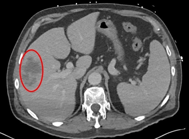
Medical Image of the Month: Hepatic Abscess Secondary to Diverticulitis
   Resulting in Sepsis
Medical Image of the Month: Metastatic Spindle Cell Carcinoma of the
   Breast
Medical Image of the Month: Perforated Gangrenous Cholecystitis


For complete imaging listings click here

Those who care for patients with pulmonary, critical care or sleep disorders rely heavily on chest radiology and pathology to determine diagnoses. The Southwest Journal of Pulmonary, Critical Care & Sleep publishes case-based articles with characteristic chest imaging and related pathology. The editor of this section will oversee and coordinate the publication of a core of the most important chest imaging topics. In doing so, they encourage the submission of unsolicited manuscripts. It cannot be overemphasized that both radiologic and pathologic images must be of excellent quality. As a rule, 600 DPI is sufficient for radiographic and pathologic images. Taking pictures of plain chest radiographs and CT scans with a digital camera is strongly discouraged. The figures should be cited in the text and numbered consecutively. The stain used for pathology specimens and magnification should be mentioned in the figure legend. Those who care for patients with pulmonary, critical care or sleep disorders rely heavily on chest radiology and pathology to determine diagnoses. The Southwest Journal of Pulmonary, Critical Care & Sleep publishes case-based articles with characteristic chest imaging and related pathology. The editor of this section will oversee and coordinate the publication of a core of the most important chest imaging topics. In doing so, they encourage the submission of unsolicited manuscripts. It cannot be overemphasized that both radiologic and pathologic images must be of excellent quality. As a rule, 600 DPI is sufficient for radiographic and pathologic images. Taking pictures of plain chest radiographs and CT scans with a digital camera is strongly discouraged. The figures should be cited in the text and numbered consecutively. The stain used for pathology specimens and magnification should be mentioned in the figure legend.

-------------------------------------------------------------------------------------------  

Monday
Nov022020

Medical Image of the Month: Buffalo Chest Identified at the Time of Lung Nodule Biopsy

Figure 1. Buffalo chest, peripheral cavitary lesions, and pneumothorax contralateral to the biopsy site. A) Outpatient CT scan showing a right pneumothorax (blue arrow) and right-sided cavitary lesion (blue arrowhead). B) Subsequent pre-procedural planning CT scan done right lateral decubitus, showing again the cavitary lesion (blue arrowhead) but now with left pneumothorax (red arrow), suggesting buffalo chest. C) Intra-procedural CT showing needle at the periphery of the cavitary lesion (blue arrowhead) and similar-sized left pneumothorax (red arrow). D) 5-minute post-procedural CT demonstrating expected alveolar hemorrhage in the site of the biopsied cavitary lesion (blue arrowhead), but worsening left pneumothorax (red arrow). E & F) Multiple peripheral left lung cavitary lesions (red arrowheads) felt to be possible culprits for the worsened pneumothorax following coughing from the right-sided biopsy.


A gentleman in his late 50s with a past medical history of squamous cell carcinoma at the base of the tongue had numerous slowly-growing pneumocyst-like lesions despite clinical remission status post surgery and chemoradiation. Biopsy of one of these lesions was recommended by a multidisciplinary tumor board.

An outpatient pre-procedural supine chest CT revealed a right pneumothorax above the lesion targeted for biopsy. A subsequent pre-procedural right lateral decubitus chest CT three weeks later demonstrated a left-sided pneumothorax, raising concern for buffalo chest. (A less likely possibility would be spontaneous resolution of the right pneumothorax and development of a new left pneumothorax in the less than 4-week interval.) Intraprocedural imaging continued to demonstrate the left-sided pneumothorax. A biopsy touch preparation of the first sample obtained did not demonstrate malignancy. Therefore, an attempt was made at obtaining another sample. However, the patient developed a brief but forceful coughing fit, resulting in the termination of the procedure after only 2 passes of a 20g needle. Post-procedural scans demonstrated expected right-sided alveolar hemorrhage near the biopsy tract but slight worsening of the contralateral pneumothorax. The patient was again scanned five minutes later for concerns of a worsening pneumothorax. CT imaging demonstrated stable right-sided alveolar hemorrhage near the biopsy tract, but also severe left sided pneumothorax with multiple peripheral cavitary lesions. A left anterior chest tube was placed and the patient was discharged a few days later. Biopsy results showed granulomatous tissue only and cultures were negative.

Buffalo chest refers to the abnormal presence of a pleuro-pleural communication in humans. This phenomenon derives its name from the fact that buffalo and similar species possess one contiguous pleural space, while humans ordinarily have two independent pleural spaces corresponding to each lung (1). This pleuro-pleural communication can develop iatrogenically, congenitally, or as a result of trauma. Cases of buffalo chest developing after median sternotomy, laparoscopic surgery and heart-lung transplantation have been described in the literature (2). It is unknown if the worsening of our patient’s left-sided pneumothorax occurred in the setting of underlying buffalo chest, with biopsy producing a right sided pneumothorax that subsequently communicated with the left pleural space. Alternatively, our patient’s episode of forceful coughing could have increased the intrathoracic pressure and caused barotrauma to one of the many peripheral cavitary lesions previously described, leading to a worsening of the left-sided pneumothorax. Cases of bilateral pneumothoraces where interpleural connections exist have been successfully treated with a unilateral chest tube (2, 3). A second chest tube may be necessary in cases where the contralateral lung fails to re-expand. Our patient was able to be discharged after placement of a unilateral chest tube (contralateral to the biopsy side), reaffirming the general recommendation to treat pneumothoraces this patient population with a unilateral pleural catheter.

Phillip Belone MS4, Jason Lee MD, and Michael Craig Larson MD PhD

University of Arizona/Banner University Medical Center-Tucson

Department of Medical Imaging

Tucson, AZ USA

References

  1. Jacobi A, Eber C, Weinberger A, Friedman SN. Bilateral Pneumothoraces after Unilateral Lung Biopsy. A Case of "Buffalo Chest"? Am J Respir Crit Care Med. 2016 Apr 15;193(8):e36. [CrossRef] [PubMed]
  2. Sawalha L, Gibbons WJ. Iatrogenic "buffalo chest" bilateral pneumothoraces following unilateral transbronchial lung biopsies in a bilateral lung transplant recipient. Respir Med Case Rep. 2015 May 16;15:57-8. [CrossRef] [PubMed]
  3. Groarke J, Breen D, O'Connell F, O'Donnell R. Bilateral pneumothorax resulting from a diagnostic thoracentesis. Eur Respir J. 2007 Nov;30(5):1018-9; diagnosis 1020. [CrossRef] [PubMed]

Cite as: Belone P, Lee J, Larson MC. Medical Image of the Month: Buffalo Chest Identified at the Time of Lung Nodule Biopsy. Southwest J Pulm Crit Care. 2020;21(5):121-3. doi: https://doi.org/10.13175/swjpcc056-20 PDF

Sunday
Nov012020

November 2020 Imaging Case of the Month: Cause and Effect?

Prasad M. Panse MD 

Clinton E. Jokerst MD 

Michael B. Gotway MD 

Department of Radiology

Mayo Clinic, Arizona

Phoenix, Arizona USA


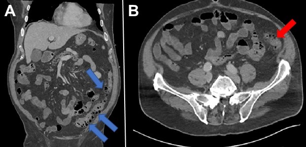
Clinical History: A 36 -year-old woman with Crohn’s disease and ulcerative colitis diagnosed approximately 1 year earlier, was initially treated with adalimumab, but later switched to prednisone and budesonide when subcutaneous nodules and migraines were attributed to this medication. Subsequently a flare of gastrointestinal symptoms prompted hospitalization with colonoscopy which showed severe pancolitis consistent with ulcerative colitis. One month following hospital discharge, the patient then presented to the Emergency Department with continued complaints of nausea, diarrhea, abdominal pain, intermittent fever (self-measured to 101º F), joint pain, and a pruritic rash all over her body. These symptoms had occurred following her hospitalization 2 months earlier. She also complained of 25 lbs. weight loss over the previous year.

In addition to prednisone and budesonide, the patient’s medications included hydroxyzine, famotidine, vitamin C, and hydrocodone-acetaminophen. The patient denies allergies and did not smoke nor use drugs.

Physical examination showed the patient to be afebrile with normal heart and respiratory rates and blood pressure = 112/75 mmHg. Her room air oxygen saturation was 99%. Her examination was remarkable for tenderness to palpation over the left > right lower quadrants with rebound tenderness and positive fecal occult blood testing. Her skin examination also showed a diffuse, pinpoint, maculopapular rash affecting her trunk as well as both the upper and lower extremities.

The patient’s complete blood count and serum chemistries showed hypokalemia=3.0 mmol/L (normal, 3.6-5.2 mmol/L), mild anemia (hemoglobin / hematocrit = 11.2 gm/dL / 34.3% [normal, 12.3-15.7 gm/dL / 37-46%]), and a minimally elevated lipase of 63 U/L (normal, 13-60 U/L). Liver and renal function testing were within normal limits.

Which of the following represents an appropriate next step for the patient’s management?

  1. Obtain gastrointestinal consult
  2. Obtain a travel history
  3. Obtain abdominal CT
  4. All of the above
  5. None of the above

Cite as: Panse PM, Jokerst CE, Gotway MB. November 2020 Imaging Case of the Month: Cause and Effect? Southwest J Pulm Crit Care. 2020;21(5):108-120. doi: https://doi.org/10.13175/swjpcc058-20 PDF