Search Journal-type in search term and press enter
Southwest Pulmonary and Critical Care Fellowships
In Memoriam
Social Media

Pulmonary

Last 50 Pulmonary Postings

(Click on title to be directed to posting, most recent listed first)

March 2025 Pulmonary Case of the Month: Interstitial Lung Disease of
   Uncertain Cause
December 2024 Pulmonary Case of the Month: Two Birds in the Bush Is
   Better than One in the Hand
Glucagon‐like Peptide-1 Agonists and Smoking Cessation: A Brief Review
September 2024 Pulmonary Case of the Month: An Ounce of Prevention
   Caused a Pound of Disease
Yield and Complications of Endobronchial Ultrasound Using the Expect
   Endobronchial Ultrasound Needle
June 2024 Pulmonary Case of the Month: A Pneumo-Colic Association
March 2024 Pulmonary Case of the Month: A Nodule of a Different Color
December 2023 Pulmonary Case of the Month: A Budding Pneumonia
September 2023 Pulmonary Case of the Month: A Bone to Pick
A Case of Progressive Bleomycin Lung Toxicity Refractory to Steroid Therapy
June 2023 Pulmonary Case of the Month: An Invisible Disease
February 2023 Pulmonary Case of the Month: SCID-ing to a Diagnosis
December 2022 Pulmonary Case of the Month: New Therapy for Mediastinal
   Disease
Kaposi Sarcoma With Bilateral Chylothorax Responsive to Octreotide
September 2022 Pulmonary Case of the Month: A Sanguinary Case
Electrotonic-Cigarette or Vaping Product Use Associated Lung Injury:
   Diagnosis of Exclusion
June 2022 Pulmonary Case of the Month: A Hard Nut to Crack
March 2022 Pulmonary Case of the Month: A Sore Back Leading to 
   Sore Lungs
Diagnostic Challenges of Acute Eosinophilic Pneumonia Post Naltrexone
Injection Presenting During The COVID-19 Pandemic
Symptomatic Improvement in Cicatricial Pemphigoid of the Trachea
   Achieved with Laser Ablation Bronchoscopy
Payer Coverage of Valley Fever Diagnostic Tests
A Summary of Outpatient Recommendations for COVID-19 Patients
   and Providers December 9, 2021
December 2021 Pulmonary Case of the Month: Interstitial Lung
   Disease with Red Knuckles
Alveolopleural Fistula In COVID-19 Treated with Bronchoscopic 
   Occlusion with a Swan-Ganz Catheter
Repeat Episodes of Massive Hemoptysis Due to an Anomalous Origin 
   of the Right Bronchial Artery in a Patient with a History
   of Coccidioidomycosis
September 2021 Pulmonary Case of the Month: A 45-Year-Old Woman with
   Multiple Lung Cysts
A Case Series of Electronic or Vaping Induced Lung Injury
June 2021 Pulmonary Case of the Month: More Than a Frog in the Throat
March 2021 Pulmonary Case of the Month: Transfer for ECMO Evaluation
Association between Spirometric Parameters and Depressive Symptoms 
   in New Mexico Uranium Workers
A Population-Based Feasibility Study of Occupation and Thoracic
   Malignancies in New Mexico
Adjunctive Effects of Oral Steroids Along with Anti-Tuberculosis Drugs
   in the Management of Cervical Lymph Node Tuberculosis
Respiratory Papillomatosis with Small Cell Carcinoma: Case Report and
   Brief Review
December 2020 Pulmonary Case of the Month: Resurrection or 
   Medical Last Rites?
Results of the SWJPCC Telemedicine Questionnaire
September 2020 Pulmonary Case of the Month: An Apeeling Example
June 2020 Pulmonary Case of the Month: Twist and Shout
Case Report: The Importance of Screening for EVALI
March 2020 Pulmonary Case of the Month: Where You Look Is 
   Important
Brief Review of Coronavirus for Healthcare Professionals February 10, 2020
December 2019 Pulmonary Case of the Month: A 56-Year-Old
   Woman with Pneumonia
Severe Respiratory Disease Associated with Vaping: A Case Report
September 2019 Pulmonary Case of the Month: An HIV Patient with
   a Fever
Adherence to Prescribed Medication and Its Association with Quality of Life
Among COPD Patients Treated at a Tertiary Care Hospital in Puducherry
    – A Cross Sectional Study
June 2019 Pulmonary Case of the Month: Try, Try Again
Update and Arizona Thoracic Society Position Statement on Stem Cell 
   Therapy for Lung Disease
March 2019 Pulmonary Case of the Month: A 59-Year-Old Woman
   with Fatigue
Co-Infection with Nocardia and Mycobacterium Avium Complex (MAC)
   in a Patient with Acquired Immunodeficiency Syndrome 
Progressive Massive Fibrosis in Workers Outside the Coal Industry: A Case 
   Series from New Mexico
December 2018 Pulmonary Case of the Month: A Young Man with
   Multiple Lung Masses
Antibiotics as Anti-inflammatories in Pulmonary Diseases
September 2018 Pulmonary Case of the Month: Lung Cysts
Infected Chylothorax: A Case Report and Review
August 2018 Pulmonary Case of the Month
July 2018 Pulmonary Case of the Month
Phrenic Nerve Injury Post Catheter Ablation for Atrial Fibrillation
Evaluating a Scoring System for Predicting Thirty-Day Hospital 
   Readmissions for Chronic Obstructive Pulmonary Disease Exacerbation
Intralobar Bronchopulmonary Sequestration: A Case and Brief Review


For complete pulmonary listings click here.

The Southwest Journal of Pulmonary and Critical Care publishes articles broadly related to pulmonary medicine including thoracic surgery, transplantation, airways disease, pediatric pulmonology, anesthesiolgy, pharmacology, nursing  and more. Manuscripts may be either basic or clinical original investigations or review articles. Potential authors of review articles are encouraged to contact the editors before submission, however, unsolicited review articles will be considered.

-------------------------------------------------------------------------------------

Monday
Aug112014

Role of Endobronchial Ultrasound in the Diagnosis and Management of Bronchogenic Cysts: Two Case Descriptions and Literature Review

Rene Franco-Elizondo MD

Soumya Patnaik MD

Kuan-Hsiang Gary Huang MD, PhD

Jorge Mora MD

Albert Einstein Medical Center

Philadelphia, Pennsylvania


Abstract

Imaging studies, such as high resolution computerized tomography (HRCT) and magnetic resonance imaging (MRI) facilitate the evaluation of mediastinal masses. However, the definite characterization of such masses can be ascertained only after tissue sampling is obtained and analyzed. Some mediastinal masses, like bronchogenic cysts, can be misdiagnosed as solid masses or lymphadenopathy in imaging studies, due to the variable densities of the cyst contents. More invasive tests, like fine needle aspiration or surgical resection of the bronchogenic cyst, may be necessary when HRCT fails to provide an initial diagnosis. We describe two such cases seen at our institution that highlight the implications of establishing a diagnosis of bronchogenic cyst with endobronchial ultrasound (EBUS) - trans-bronchial needle aspiration (TBNA) and discuss the possible therapeutic utility of EBUS-TBNA in select patients with bronchogenic cysts.


Abbreviation List

BAL - Bronchoalveolar lavage

CNS - Coagulase-negative Staphylococcus

CT – Computed tomography

EBUS - Endobronchial ultrasound

EUS – Endoscopic ultrasound

FOB - Fiberoptic bronchoscopy

HRCT - High resolution computerized tomography

MRI - Magnetic resonance imaging

RUL – Right upper lobe

TBNA - Trans-bronchial needle aspiration

VATS - Video-assisted thoracoscopic surgery

Introduction

Modern imaging, particularly high-resolution computed tomography (HRCT) and magnetic resonance imaging facilitate the evaluation of mediastinal masses. However, definite characterization is possible only after tissue sampling is obtained, typically through fine-needle aspiration or surgical resection. Herein, we report two cases of patients with mediastinal masses, where HRCT failed to provide a diagnosis. Bronchogenic cysts in both patients were ultimately diagnosed by endobronchial ultrasound (EBUS) and trans-bronchial needle aspiration (TBNA). The implications of establishing a diagnosis of bronchogenic cyst via EBUS-TBNA and therapeutic approaches are discussed.

Case 1

A 68-year-old African American woman with hypertension, diabetes mellitus type 2 and end-stage renal disease, on home hemodialysis, presented to the hospital with central stabbing chest pain, radiating to the back, accompanied by shortness of breath. An initial HRCT chest performed to rule out aortic dissection revealed a large subcarinal mass, measuring 2.3 cm x 6.5 cm x 3.8 cm (AP x transverse x height), that splayed the carina, exerting mass effect on the esophagus, raising suspicion of malignancy (Figure 1).

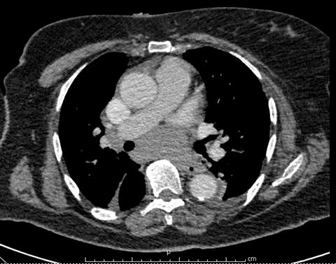
Figure 1. Subcarinal mass. Density ranged from 25-80 Hounsfield Units.

A separate 2.2 cm x 1.7 cm right paratracheal mass, mediastinal lymphadenopathy and many small prevascular lymph nodes were noted. These clinical and imaging findings were concerning for possible lymphoma.

A fiberoptic bronchoscopy (FOB), followed by blind TBNA of the subcarinal space using a Wang needle was attempted. Both, bronchoalveolar lavage (BAL) and TBNA were unrevealing. The patient was found to have persistent coagulase-negative Staphylococcus (CNS) bacteremia, with the first blood cultures being positive at the time of admission. A thorough evaluation, which included an echocardiogram and abdominal HRCT, failed to reveal a source of bacteremia, which was ultimately thought to be related to hemodialysis. Arrangements for outpatient EBUS evaluation of the mediastinal mass and lymphadenopathy were made and the patient was discharged.

A week later, she was readmitted for hypertensive emergency. EBUS was performed during this hospitalization and a cystic mass with heterogeneous-containing material was detected in the subcarinal space (Figure 2).

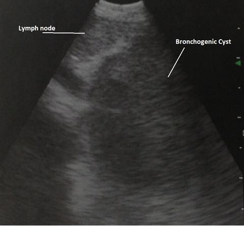
Figure 2 EBUS image of bronchogenic cyst and adjacent subcarinal lymph node.

The needle aspirate was sent for histological analysis and culture, but it was not possible to drain this cystic structure. Cytopathologic analysis showed bronchial and ciliated cells with abundant mucoid material and a diagnosis of bronchogenic cyst was made. Interestingly, the cultures from the aspirated material grew CNS. The patient was discharged with plans for a video-assisted thoracoscopic surgery (VATS) resection of the bronchogenic cyst as an outpatient.

Five days later, the patient was readmitted with symptoms that were concerning for sepsis, and was thus re-started on broad-spectrum antibiotics. She was found to have Enterobacter intermedius bacteremia. She subsequently underwent VATS, with direct aspiration of the bronchogenic cyst. A resection was not performed due to technical difficulties encountered during VATS. Purulent fluid was retrieved from the cyst and Enterobacter intermedius was identified upon analysis and culture of the cyst content. The patient had no further episodes of bacteremia after eight months of follow-up.

Case 2

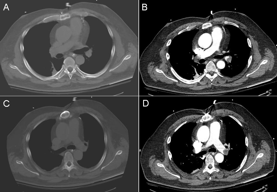
A 43-year-old woman without significant past medical history was referred to our institute, for evaluation of a pretracheal lymph node seen on a chest HRCT (Figure 3) done for evaluation of new onset dyspnea and wheezing. Upon auscultation, a localized wheeze was noted with deep inspiration in the right upper chest. Her physical exam was otherwise unremarkable.

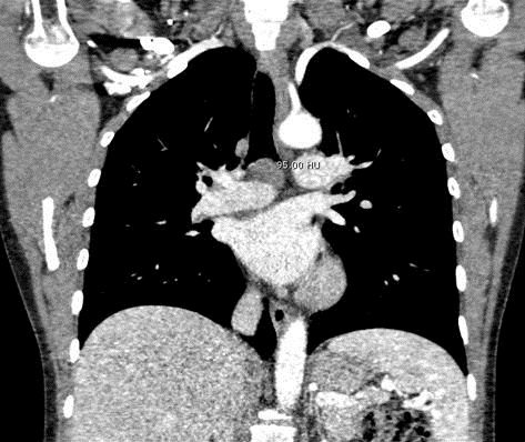
Figure 3. Chest CT showing subcarinal lymphadenopathy and mass. Density of mass 9-95 Hounsfield units.

A bronchoscopic exam with EBUS evaluation of lymphadenopathy was scheduled. On FOB, the patient was found to have an incidental endobronchial mass occluding the anterior segment of the right upper lobe. EBUS exam revealed an enlarged subcarinal lymph node (8 mm) with an adjacent cystic space containing homogenous hypoechoic material (Figure 4).

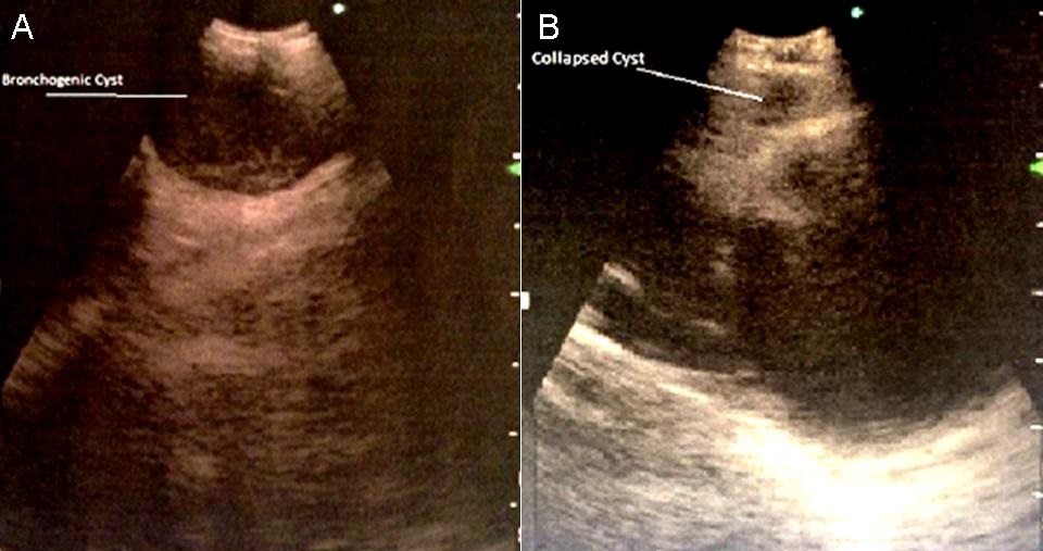
Figure 4. EBUS of bronchogenic cyst. A) Cyst prior to aspiration. B) Collapsed cystic cavity with enlarged lymph node now visible.

Both the lymph node and cystic space were sampled. Ten mL of serous fluid was aspirated from the cystic space, resulting in obliteration of the cavity, as visualized on the ultrasound (Figure 5).

Figure 5.Serous aspirate from cystic cavity.

Full mediastinal staging was done, and only station 11R lymph nodes were found to be enlarged and were sampled. Endobronchial biopsies, brushings and BAL were obtained from RUL endobronchial lesion. The patient was discharged home on empiric antibiotics (amoxicillin/clavulanate) for aspirated bronchogenic cyst. Subsequent fluid analysis revealed abundant macrophages and lymphocytes, consistent with cystic fluid content. Cultures of the fluid were positive for Streptococcus viridians. Lymph node sampling failed to reveal any evidence of malignancy. Interestingly, endobronchial mass biopsies, brushings and fluid cytology also failed to show evidence of malignancy. Only reactive inflammatory cells and benign bronchial elements were detected. The patient was continued on antibiotics for ten days without any evidence of infection.

A repeat bronchoscopy was performed to re-sample the endobronchial lesion. Benign elements were confirmed on the repeat biopsy. Follow up imaging has not been performed to evaluate fluid re-accumulation, since the patient has remained asymptomatic for two months.

Discussion

Mediastinal bronchogenic cysts are congenital anomalies of tracheobronchial origin; they are believed to be a result of an abnormal budding process during the development of foregut. They are often asymptomatic at presentation but can become symptomatic in 30% to 80% of cases due to infection or other complications like compressive efforts (1).

These cysts, being lined by secretary respiratory epithelium, consist of fluid of water density; however, the amount of proteinous mucus and calcium oxalate crystals in them can vary, affecting the imaging features on CT/MRI. A chest CT may reveal spherical masses with water or soft–tissue attenuation. A chest CT may misdiagnose them as soft-tissue masses in about 43% of patients. High attenuation on a chest CT can be a result of calcium oxalate or protein content, or can be due to infection of the cyst content (2, 3).

Due to the variable density in the cyst’s content, bronchogenic cysts can be misdiagnosed as masses or lymphadenopathy on non-invasive testing, as noted in our patients. EBUS can be of great help in diagnosing these lesions. Ultrasound provides an excellent delineation between tissues of different densities, and the absence of flow with color Doppler allows for differentiation from vascular structures. Ultrasonography allows a better delineation of cystic lesions and characterization of their contents (e.g. hypoechoic, isoechoic, heterogeneous, etc.), thereby providing useful diagnostic information. Needle aspiration of cyst contents can bring about not only cytological confirmation of the diagnosis, but also identification of complications such as infected bronchogenic cysts.

Our cases highlight the usefulness of EBUS in the diagnosis of bronchogenic cysts. In the first case, the diagnosis of bronchogenic cyst was made only after EBUS imaging and content aspiration were obtained, despite the initial chest HRCT specifically done to evaluate this mass. In the second case, EBUS imaging established the diagnosis in the absence of any suggestive findings on the HRCT.

The treatment of choice remains the complete surgical resection of the secreting mucosal lining, particularly in complicated cysts (11, 12). However, some authors have reported cases of successful treatment of bronchogenic cysts with EBUS-guided aspiration (4-8). In one case, a patient was followed up for eighteen months without evidence of recurrence (8). The rationale behind this approach is that complete drainage of the cyst obliterates the cyst cavity and prevents further fluid re-accumulation. In our first case, though complete drainage was not achieved with EBUS due to its thick mucoid content, aspiration of the cyst by VATS resulted in resolution without fluid re-accumulation. In our second case, resolution of the cyst was achieved via EBUS-TBNA drainage. These cases underscore the usefulness of aspiration of bronchogenic cysts as an alternative therapeutic approach to surgery in certain scenarios.

Contrary to the above mentioned cases, other case reports have pointed out life-threatening complications after bronchogenic cyst drainage with EBUS-guided FNA, such as pneumonia (9) or purulent pericardial effusion (10). As mentioned elsewhere, empiric antibiotic therapy should be given when a cystic lesion is drained via EBUS-TBNA (13). It should be noted, however, that in some of these case reports, infection post-EBUS-TBNA occurred despite giving empiric antibiotics (9), as in our first case.

The risk of infection should be underscored, as evidenced by the first case; particularly the less frequently reported possibility of bronchogenic cyst infection from bacteremia. The initial EBUS-TBNA cyst aspirate grew CNS, similarly to the blood cultures that were obtained prior to the blind TBNA sample of the mediastinal lesion. This suggests that the contamination of the cyst content could have been due to seeding from CNS bacteremia. However, the final VATS aspirate of the cyst grew Enterecoccus intermedius, which was likely to have been introduced by the EBUS-TBNA at the time of diagnosis. This infection occurred despite the use of antibiotics before and after the procedure. In this regard, the available literature is scant. In a study conducted by Steinfort et al. (14), incidence of bacteremia after EBUS-TBNA was found to be 7%, comparable to reported incidence of bacteremia from regular FOB. It is important to note that although none of these patients experienced clinical signs of infection, none of the biopsies were taken from cystic structures. Data evaluating EBUS-TBNA of mediastinal cystic lesions is conflicting. In a report of 22 patients undergoing EUS-TBNA of suspected mediastinal cyst and receiving periprocedural antibiotics, no infectious complications were found (15). However, several case reports of serious infectious complications after EBUS-TBNA have also been published (16).

Conclusion

Diagnosis of bronchogenic cysts cannot always be made with commonly used chest-imaging modalities such as X-ray or CT. EBUS has proven to be a useful diagnostic tool in the evaluation of some mediastinal masses. Although surgical resection remains the treatment of choice, complete aspiration, by VATS or EBUS, can be a successful therapeutic alternative in patients who are not candidates for surgery. However, the risks should be carefully assessed in each patient, with particular awareness of potential infectious complications. When this approach is taken, empiric antibiotics are recommended.

References

  1. St-Georges R, Deslauriers J, Duranceau A, Vaillancourt R, Deschamps C, Beauchamp G, Pagé A, Brisson J. Clinical spectrum of bronchogenic cysts of mediastinum and lung in adult. Ann Thorac Surg. 1991;52:6-13. [CrossRef] [PubMed]
  2. Mc Adams HP, Kirejczyk WM, Rosado-de-Christenson ML, Matsumoto S. Bronchogenic cyst: imaging features with clinical and histopathological correlation. Radiology. 2000;217:441-6. [CrossRef] [PubMed] 
  3. Patel SR, Meeker DP, Biscotti CV, Kirby TJ, Rice TW. Presentation and management of bronchogenic cysts in adult. Chest 1994;106:79-85. [CrossRef] [PubMed] 
  4. Aragaki-Nakahodo AA, Guitron-Roig J, Eschenbacher W, Benzaquen S, Cudzilo C. Endobronchial ultrasound-guided needle aspiration of a bronchogenic cyst to liberate from mechanical ventilation: case report and literature review. J Bronchology Interv Pulmonol. 2013;20(2):152-4. [CrossRef] [PubMed]
  5. Meseguer SM, Franco-Serrano J. Drainage of a mediastinal cyst by endobronchial ultrasound-guided needle aspiration. Arch Bronconeumol. 2010;46(4):207-8. [CrossRef]  [PubMed]
  6. Dhand S and Krimsky W. Bronchogenic cyst treated by endobronchial ultrasound drainage. Thorax. 2008;63(4):386. [CrossRef] [PubMed]
  7. Galluccio G, Lucantoni G. Mediastinal bronchogenic cyst's recurrence treated with EBUS-FNA with a long-term follow-up. Eur J Cardiothoracic Surg. 2006; 29(4):627-9. [CrossRef] [PubMed]
  8. Casal RF, Jimenez CA, Mehran RJ, Eapen GA, Ost D, Sarkiss M, Morice RC. Infected mediastinal bronchogenic cyst successfully treated by endobronchial ultrasound-guided fine-needle aspiration. Ann Thorac Surg. 2010; 90(4):e52-3. [CrossRef] [PubMed]
  9. Hong G, Song J, Lee KJ, Jeon K, Koh WJ, Suh GY, Chung MP, Kim H, Kwon OJ, Um SW. Bronchogenic cyst rupture and pneumonia after endobronchial ultrasound-guided transbronchial needle aspiration: a case report. Tuberc Respir Dis (Seoul). 2013;74(4):177-80. [CrossRef] [PubMed] 
  10. Gamrekeli A, Kalweit G, Schäfer H, Huwer H. Infection of a Bronchogenic cyst after ultrasonography-guided fine needle aspiration. Ann Thorac Surg. 2013;95(6):2154-5. [CrossRef] [PubMed]
  11. Cioffi U, Bonavina L, De Simone M, Santambrogio L, Pavoni G, Testori A, Peracchia A. Presentation and surgical management of bronchogenic and esophageal duplication cysts in adults. Chest. 1998;113(6):1492-6. [CrossRef] [PubMed] 
  12. Anantham D, Phua GC, Low SY, Koh MS. Role of endobronchial ultrasound in the diagnosis of bronchogenic cysts. Diagn Ther Endosc. 2011, 2011:468237. [CrossRef] [PubMed]
  13. Haas AR. Infectious complications from full extension endobronchial ultrasound transbronchial needle aspiration. Eur Respir J. 2009;33(4):935-8. [CrossRef] [PubMed]
  14. Steinfort DP, Johnson DF, Irving L.B. Incidence of bacteraemia following endobronchial ultrasound-guided transbronchial needle aspiration. Eur Respir J. 2010:36(1):28-32. [CrossRef] [PubMed] 
  15. Fazel A, Moezardalan K, Varadarajulu S, Draganov P, Eloubeidi MA. The utility and the safety of EUS-guided FNA in the evaluation of duplication cysts.GastrointestEndosc. 2005; 62(4):575-80. [CrossRef] [PubMed]
  16. Jenssen C, Alvarez-Sánchez M. V., Napoléon B and Faiss S. Diagnostic endoscopic ultrasonography: assessment of safety and prevention of complications. World J Gastroenterol. 2012;18(34):4659–76. [CrossRef] [PubMed]

Reference as: Franco-Elizondo R, Patnaik S, Huang K-H G, Mora J. Role of endobronchial ultrasound in the diagnosis and management of bronchogenic cysts: two case descriptions and literature review. Southwest J Pulm Crit Care. 2014;9(2):115-22. doi: http://dx.doi.org/10.13175/swjpcc096-14 PDF

Tuesday
Aug052014

Azathioprine Associated Acute Respiratory Distress Syndrome: Case Report and Literature Review

Dmitriy Scherbak, D.O.

Ruth Wyckoff, M.D.

Clement Singarajah, M.D.


Phoenix VA Healthcare System

Phoenix, AZ


Abstract

A 58-year-old Caucasian man treated with azathioprine to prevent rejection of an orthotopic liver transplant, presented to the Carl Hayden VA Medical Center with rapid respiratory decline and appeared septic. He required urgent intubation, mechanical ventilator support and empiric antibiotics. His clinical picture and imaging studies were consistent with acute respiratory distress syndrome; however, extensive infectious work up failed to reveal an offending organism. Review of his current medications implicated azathioprine and upon discontinuation of this agent, the patient made a rapid recovery. He was subsequently extubated, transferred out of the ICU and soon discharged home in good health.

Prescribed for organ transplant rejection and a wide array of autoimmune diseases, azathioprine has been rarely correlated with pneumonitis and rapid respiratory failure. No reported cases were found in which azathioprine was used to treat liver transplant rejection and associated with development of the adult respiratory distress syndrome (ARDS). However, there have been ARDS cases in which azathioprine was used for other purposes. We review all the available cases of azathioprine associated ARDS. The patients in these reports had similar clinical symptoms on presentation as our patient: hypoxia, febrile episodes and rapid development of ARDS with no infectious etiology. Most notable is the rapid resolution of ARDS after discontinuation of azathioprine.

Although azathioprine toxicity related respiratory failure is rare, this correlation should still be considered in the differential for immunosuppressed patients presenting with rapid pulmonary decline. Further studies are needed and warranted to better correlate this connection, but it is imperative to recognize that the relationship exists.

Introduction

Since its first use in 1961, azathioprine (a derivative of 6-mercaptopurine) has been used as a steroid sparing immunosuppressive agent in numerous disorders including prevention of graft rejection for solid organ transplantation (1-2). Azathioprine side effects are commonly gastrointestinal complaints such as nausea and vomiting, occurring in ~19% of patients. Laboratory abnormalities such as leukopenia are also common (17%) with thrombocytopenia and anemia being less common (3-4%) (3). Hepatotoxicity has been reported as well. Pulmonary toxicity is not usually noted as a side effect (1). Sixteen cases have been reported in the literature implicating azathioprine with pulmonary toxicity (1-2, 4-12). In 10 of these cases, the patient developed acute respiratory distress syndrome (ARDS) (1,2,6,8,9,11).

Pulmonary infections have been the leading cause of complications in immunosuppressed recipients of solid organs (13). Therefore, when a patient presents with respiratory distress, an abnormal chest x-ray and fevers, such infections are high on the differential, but the possibility of lung injury resulting from the immunosuppressive agent is often overlooked (1). We present a case of azathioprine induced ARDS in a liver transplant recipient and review the available ARDS cases associated with azathioprine use.

Case Report

We present a 58-year-old white man with a past medical history of end-stage liver disease due to hepatitis C cirrhosis and hepatocellular carcinoma who received an orthotopic liver transplant (OLT) 9 months prior to presentation. He was being treated with azathioprine 150mg daily and tacrolimus 1.5 mg daily to prevent rejection. He presented to the emergency department 9 months after his transplant with shortness of breath and increasing hypoxia. He was admitted to the intensive care unit where he developed respiratory failure that night requiring intubation and ventilator support. He had fevers as high as 105.1⁰F. He had pancytopenia with white blood cell count (WBC) 2.3 thousand cells at presentation, hemoglobin (HGB) 9.8 g/dL and platelets (PLT) 119 thousand cells.

Chest x-ray showed bilateral patchy pulmonary infiltrates. CT of the chest was done as well showing bilateral ground glass opacities and diffuse scattered pulmonary consolidations (Figure 1).

Figure 1. Representative images from chest CT with contrast done on admission showing diffuse ground glass opacities and scattered pulmonary consolidations.

Since he was immunosuppressed he was started on empiric antibiotic coverage with vancomycin, levofloxacin, pipercillin/tazobactam, gancyclovir and fluconazole. Trimethoprim-sulfamethoxazole was added on day 2 of hospitalization. A bronchoscopy with bronchial alveolar lavage (BAL) was done prior to antibiotics. Cell count and differential showed 160 white blood cells, 11% segmented neutrophils and 3% eosinophils, the other 86% of cells were pulmonary macrophages/monocytes and reactive respiratory epithelial cells. No organisms or evidence of malignancy were seen. BAL cultures showed no growth on bacterial, viral, acid fast or mycology cultures. Influenza A and B and a pneumocystis smear were also negative. Blood cultures were taken twice during the patient’s hospitalization during febrile episodes and showed no growth both times in two sets of cultures. On day 6 of hospitalization anti-microbial therapy was discontinued.

The patient’s clinical status continued to deteriorate. Chest x-rays continued to show increasing bilateral pulmonary infiltrates (Figure 2).

Figure 2. Chest x-ray at worst (hospital day 8) showing worsening bilateral pulmonary infiltrates.

The diagnosis of acute respiratory distress syndrome (ARDS) was established. His ventilator settings followed the NHLBI ARDS Network protocol, and on day 6 he was even placed in a prone position. On day 7 of hospitalization his white blood cell count dropped to a nadir of 0.5 thousand cells, hemoglobin dropped to 6.5 g/dL and platelets down to 69 thousand cells). Azathioprine was discontinued due to the pancytopenia and due to finding a few case reports in which it was implicated in ARDS. Within 3 days of azathioprine discontinuation (day 10 of hospitalization), the patient’s chest x-rays and pulmonary function had dramatically improved and he was successfully extubated by the fifth day of azathioprine being withdrawn (day 12 of hospitalization). Daily chest x-rays showed continued resolution of infiltrates (Figure 3).

Figure 3. Chest x-ray from hospital day 15 showing dramatic improvement of infiltrates after azathioprine discontinuation.

He improved rapidly and was discharged from the ICU on day 17 and discharged home from the hospital on day 18 with complete resolution of his pulmonary symptoms. His azathioprine was not restarted but he resumed tacrolimus for immunosuppression. Six months after admission, the patient was in good health with no clinical symptoms.

Discussion

Azathioprine is a nitroimidazole derivative of 6-mercaptopurine (4). It was first used in 1961 and has since become a common medication for treatment of numerous auto-immune disorders and as an immunosuppressant in transplant recipients (1). It has been described to have several reversible dose dependent side effects including bone marrow suppression, hepatotoxicity, anorexia, nausea and vomiting (4). Hypersensitivity reactions have also been described and include fevers, rigors, arthralgia, myalgia, cutaneous reactions, headaches, interstitial nephritis, pancreatitis, dyspnea, cough and pneumonitis (1-4, 6).

In our case the patient developed pneumonitis and ARDS which resolved rapidly after the discontinuation of azathioprine. A review of the literature using broad search terms in OVID, Pub-Med and Google Scholar revealed only 10 articles constituting 16 cases of pulmonary toxicity linked to azathioprine. Detailed analysis showed only 5 reported cases of ARDS linked to azathioprine toxicity (2,6,8,9,11), and a single case series of 7 cases of which 2 also have an infectious etiology (1). Data from these cases are summarized on table 1.

Table 1. Cases of Azathioprine induced ARDS in the literature.

The four remaining articles not appearing in table 1 were excluded because they either represented an immediate hypersensitivity reaction to azathioprine or had infectious pneumonitis which could have contributed to the development of ARDS (4,5,10,12).

Neither our case nor those in the literature contain irrefutable proof that azathioprine was directly responsible for lung injury. However, the similarities between the cases in which the patient survived lead us to conclude that azathioprine is involved in this adverse reaction. First, all 8 cases in which the patient survived show a rapid improvement within one to two weeks after discontinuation of azathioprine. Second, all of these patients present in the same way with hypoxia, pulmonary infiltrates, and fevers. Third, none of the cases show any other possible causes and the ones that go to biopsy have non-specific findings (UIP or diffuse alveolar damage) (1,2,6,7,11). These observations are circumstantial, but the diagnosis of drug-induced pulmonary toxicity is usually based on clinical history of drug exposure and the absence of other known causative agents. Additionally, diffuse interstitial pulmonary disease is the most common form of lung pathology caused by drugs (1,14).

Leukopenia or pancytopenia were present in our case as well as 4 of the 10 reported cases (6,8,9,11). No other side effects from azathioprine were reported in any of the cases. Therefore ARDS is likely a unique effect and unrelated to other potential side effects of azathioprine. The dose of azathioprine was widely variable in the known cases (25-150mg daily) leading us to believe that the development of ARDS is not dose-dependent. All of the cases had patients who had been on azathioprine for months (years in one case) prior to developing pneumonitis or ARDS, leading us to speculate that ARDS is not an acute hypersensitivity. It may be that ARDS development is a function of dose effect over time.

Although there are very few reported cases, It is possible that azathioprine induced lung injury is more common than it appears. When an immunosuppressed patient presents with respiratory distress, some form of infectious etiology is usually involved and the immunosuppressants are often discontinued (1). It is possible that in some of these cases azathioprine itself is the cause or may at least contribute to the development of ARDS. We believe it is important that azathioprine lung toxicity be included in the differential for ARDS causes because prompt discontinuation of azathioprine has led to rapid recovery and good outcome in 8 of the 10 known cases (1,2,6,8,9,11).

Acknowledgments

Sarah Waybright, Pharm.D. and Lindsay Kittler, Pharm.D. The clinical pharmacists who noted case reports of azathioprine causing pulmonary toxicity and recommended it’s discontinuation in our patient.

References

  1. Bedrossian CW, Sussman J, Conklin RH, Kahan B. Azathioprine-associated interstitial pneumonitis. Am J of Clin Pathol. 1984;82(2):148-54. [PubMed]
  2. Weisenburger DD. Interstitial pneumonitis associated with azathioprine therapy. Am J of Clin Pathol. 1978;69(2):181-5. [PubMed]
  3. Whisnant JK, Pelkey J. Rheumatoid arthritis: treatment with azathioprine (IMURAN (R)). Clinical side-effects and laboratory abnormalities. Ann Rheum Dis. 1982;41:44-47. [CrossRef] [PubMed]
  4. Stetter M, Schmidl M, Krapf R. Azathioprine hypersensitivity mimicking Goodpasture's syndrome. Am J of Kidney Dis. 1994;23(6):874-7. [CrossRef] [PubMed]
  5. Krowka MJ, Breuer RI, Kehoe TJ. Azathioprine-associated pulmonary dysfunction. Chest. 1983;83(4):696-8. [CrossRef] [PubMed]
  6. Rubin G, Baume P, Vandenberg R. Azathioprine and acute restrictive lung disease. Aust N Z J Med. 1972 Aug;2(3):272-4. [CrossRef] [PubMed]
  7. Bidinger JJ, Sky K, Battafarano DF. Henning JS. The cutaneous and systemic manifestations of azathioprine hypersensitivity syndrome. [Review] J Am Acad Dermatol. 2011;65(1):184-91. [CrossRef] [PubMed]
  8. Carmichael DJS, Hamilton DV, Evans DB, Stovin PGI, Calne RY. Interstitial pneumonitis secondary to azathioprine in a renal transplant patient. Thorax. 1983;38:951-952. [CrossRef] [PubMed]
  9. Perreaux F, Delphine Z, Capron F, Trioche P, Odievre M, Labrune P. Azathioprine-induced lung toxicity and efficacy of cyclosporine a in a young girl with type 2 autoimmune hepatitis. J Ped Gastroenterol Nutr. 2000;31:190-192. [CrossRef]
  10. Ananthakrishnan AN, Attila T, Otterson MF, Lipchik RJ, Massey BT, Komorowski RA, Binion DG. Severe pulmonary toxicity after azathioprine/6-mercatopurine initiation for treatment of inflammatory bowel disease. J Clin Gastroenterol. 2007;41(7):682-688. [CrossRef] [PubMed]
  11. Brown AL, Corris PA, Ashcroft T, Wilkinson R. Azathioprine-related interstitial pneumonitis in a renal transplant recipient. Nephrol Dial Transplant. 1992;7:362-364. [PubMed]
  12. Frost J, Carion G, Mazer J. A case of azathioprine hypersensitivity syndrome, acute respiratory distress syndrome, and shock. Crit Care Med Suppl. 2011;32(12):926.
  13. De Gasperi A, Feltracco P, Ceravola E, Mazza E. Pulmonary complications in patients receiving a solid-organ transplant. Curr Opin Crit Care. 2014;20(4):411-419. [CrossRef] [PubMed]
  14. Camus P, Fanton A, Bonniaud P, Camus C, Foucher P. Interstitial lung disease induced by drugs and radiation. Respir. 2004;71:01-326. [CrossRef] [PubMed]

Reference as: Scherbak D, Wyckoff R, Singarajah C. Azathioprine associated acute respiratory distress syndrome: case report and literature review. Southwest J Pulm Crit Care. 2014;9(2):94-100. doi: http://dx.doi.org/10.13175/swjpcc087-14 PDF


Friday
Aug012014

August 2014 Pulmonary Case of the Month: A Physician's Job is Never Done

Elijah Poulos, MD*

Kristine Saunders, MD


Pulmonary and Critical Care Medicine*

Department of Pathology

Phoenix VA Medical Center

Phoenix, AZ


History of Present Illness

A 75-year-old man presented with recurrent minimally productive cough, dyspnea, fatigue, low-grade fevers, and weight loss in November 2013. The patient had been treated twice as an outpatient with antibiotics in the previous 6 weeks for pneumonia.

PMH, FH, SH

The patient has a history of obstructive sleep apnea but is not compliant with his prescribed continuous positive airway pressure. He also as a history of obesity, dyslipidemia, and peripheral vascular disease.

There is no significant family history.  

He is a retired brick layer with a 50 pack-year smoking history but quit a few weeks prior to admission.  He drinks a case of beer/week.

Physical Examination

VS stable. There were no significant findings on physical examination.

Radiography

A chest radiograph (Figure 1) was performed.

Figure 1. Admission PA (Panel A) and lateral (Panel B) chest radiograph.

What should be done next? (Click on the correct answer to proceed to the next panel)

  1. Bronchoscopy with bronchoalveolar lavage
  2. Bronchoscopy with transbronchial biopsy
  3. Needle biopsy
  4. Thoracentesis
  5. Video-assisted thorascopic surgery (VATS)

Reference as: Poulos E, Saunders K. August 2014 pulmonary case of the month: a physician's job is never done. Southwest J Pulm Crit Care. 2014;9(2):59-67. doi: http://dx.doi.org/10.13175/swjpcc098-14 PDF


Tuesday
Jul012014

July 2014 Pulmonary Case of the Month: Where Did It Come From?

Colin B. Fitterer, MD

James M. Parish, MD


Mayo Clinic Arizona

Scottsdale, AZ


History of Present Illness

A 67 year old man presented with worsening cough and shortness of breath. He has a history of metastatic colon cancer first diagnosed in 2010. He was treated with radiation and chemotherapy (FOLFOX) but unfortunately developed new pulmonary nodules in October, 2013 which were metastatic colon cancer on biopsy. In February 2014 he developed a right parietal brain mass which was resected. Thoracic CT scan at that time showed progression of the pulmonary nodules. He has also noted a 30 pound weight loss over the past 6 months and an enlarging right supraclavicular lymph node.

PMH, FH, SH

In addition to the colon cancer, he has previous diagnoses of type 2 diabetes mellitus, hypertension, allergic rhinitis, and vitamin D deficiency. He is married and a recently retired railroad engineer. He has no history of tobacco use. There is a positive family history of lung cancer but no colon cancer.

Physical Examination

Vital Signs:  Temperature 36.8, pulse 98, respirations 18, blood pressure 144/70, SpO2 91% on 3 L via nasal cannula.

Pertinent findings include:

  • A large firm and fixed right supraclavicular lymph node that is nonpainful on palpation.
  • Diminished breath sounds across all right posterior lung fields with dullness to percussion. 
  • Palpable liver edge is palpable approximately 2cm below the right costal margin.

Laboratory Analysis

Admission laboratory values include a hemoglobin of 11.1 g/dL but with a normal white blood cell count and platelet count. Electrolytes, blood urea nitrogen, creatinine, and liver enzymes were all within normal limits.  Serum chemistries are within normal limits.

Radiography

A chest x-ray (Figure 1A) and chest CT (Figure 1B) were performed.


Figure 1. Admission AP (Panel A) and representative image from the thoracic CT scan (Panel B).

Which of the following is the best interpretation of the radiographic findings? (Click on the correct answer to proceed to the next panel)

  1. Large right pleural effusion
  2. Right lung atelectasis
  3. Right lung pneumonia
  4. Right lung pulmonary edema
  5. None of the above

Reference as: Fitterer CB, Parish JM. July 2014 pulmonary case of the month: where did it come from? Southwest J Pulm Crit Care. 2014;8(6):1-7. doi: http://dx.doi.org/10.13175/swjpcc080-14 PDF


Sunday
Jun012014

June 2014 Pulmonary Case of the Month: "Petrified"

Steven W. Purtle, MD

University of Colorado Hospital, Denver, CO

steven.purtle@ucdenver.edu


History of Present Illness

A 52 year old expatriated Iraqi man presents to pulmonary clinic with complaints of chronic dyspnea. While a young man living in Iraq, he had been disqualified from service in the Iraqi Air Force after a screening chest x-ray was found to be abnormal. He had no respiratory symptoms at the time of his disqualification, and he remained asymptomatic for the next twenty five years. Beginning five years ago, he had an insidious onset of breathlessness and exertional intolerance. Over the past several years, he has developed diffuse pleuritic chest pain, non-productive cough, and fatigue. He denies any fevers, chills, night sweats, arthralgias, rash, or visual symptoms. After moving to Denver, Colorado three years ago, he has developed a continuous oxygen requirement of two liters per minute.

PMH, FH, SH

He has no significant past medical or family history. While living in Iraq, he worked as a photographer, but he is currently unemployed. He is a lifelong non-smoker and uses no illicit drugs. He has never had any pets. He denies any exposure to inorganic dusts.

Medications

None

Physical Examination

Physical examination reveals a thin, middle-aged man in no acute distress. Vital signs were notable for an oxygen saturation of 90% on 2 liters per minute of supplemental oxygen. Pulmonary examination was notable for fine inspiratory crackles heard best at the bilateral bases. There was no clubbing or peripheral edema. The remainder of his physical examination was unremarkable.

Laboratory Analysis

Serum chemistries are within normal limits. Complete blood count shows a normal white blood cell count, hematocrit, and platelet count.

Pulmonary Function Tests

Pulmonary functions tests are shown in Figure 1.

Radiography

A chest x-ray (Figure 2) and chest CT (Figures 3 and 4) were performed.

Figure 2. Admission AP (Panel A) and lateral (Panel B) chest x-ray.


Figure 3. Static thoracic CT images displayed in lung windows (Panels A-D) and soft tissue windows (Panels E-H).


Figure 4. Movies of thoracic CT scan in lung windows (Panel A, top) and soft tissue windows (Panel B, bottom).


Which of the following features best describes the pattern seen on the patient’s chest CT? (Click on the correct answer to proceed to the next panel)

  1. Diffuse microcalcifications
  2. Honeycombing
  3. Mosaicism
  4. Patchy ground glass opacifications
  5. Pleural plaques

Reference as: Purtle SW. June 2014 pulmonary case of the month: "petrified". Southwest J Pulm Crit Care. 2014;8(6):299-304. doi: http://dx.doi.org/10.13175/swjpcc069-14 PDF