Search Journal-type in search term and press enter
Southwest Pulmonary and Critical Care Fellowships
In Memoriam

Pulmonary

Last 50 Pulmonary Postings

(Click on title to be directed to posting, most recent listed first)

March 2024 Pulmonary Case of the Month: A Nodule of a Different Color
December 2023 Pulmonary Case of the Month: A Budding Pneumonia
September 2023 Pulmonary Case of the Month: A Bone to Pick
A Case of Progressive Bleomycin Lung Toxicity Refractory to Steroid Therapy
June 2023 Pulmonary Case of the Month: An Invisible Disease
February 2023 Pulmonary Case of the Month: SCID-ing to a Diagnosis
December 2022 Pulmonary Case of the Month: New Therapy for Mediastinal
   Disease
Kaposi Sarcoma With Bilateral Chylothorax Responsive to Octreotide
September 2022 Pulmonary Case of the Month: A Sanguinary Case
Electrotonic-Cigarette or Vaping Product Use Associated Lung Injury:
   Diagnosis of Exclusion
June 2022 Pulmonary Case of the Month: A Hard Nut to Crack
March 2022 Pulmonary Case of the Month: A Sore Back Leading to 
   Sore Lungs
Diagnostic Challenges of Acute Eosinophilic Pneumonia Post Naltrexone
   Injection Presenting During The COVID-19 Pandemic
Symptomatic Improvement in Cicatricial Pemphigoid of the Trachea
   Achieved with Laser Ablation Bronchoscopy
Payer Coverage of Valley Fever Diagnostic Tests
A Summary of Outpatient Recommendations for COVID-19 Patients
   and Providers December 9, 2021
December 2021 Pulmonary Case of the Month: Interstitial Lung
   Disease with Red Knuckles
Alveolopleural Fistula In COVID-19 Treated with Bronchoscopic
   Occlusion with a Swan-Ganz Catheter
Repeat Episodes of Massive Hemoptysis Due to an Anomalous Origin
   of the Right Bronchial Artery in a Patient with a History
   of Coccidioidomycosis
September 2021 Pulmonary Case of the Month: A 45-Year-Old Woman with
   Multiple Lung Cysts
A Case Series of Electronic or Vaping Induced Lung Injury
June 2021 Pulmonary Case of the Month: More Than a Frog in the Throat
March 2021 Pulmonary Case of the Month: Transfer for ECMO Evaluation
Association between Spirometric Parameters and Depressive Symptoms 
   in New Mexico Uranium Workers
A Population-Based Feasibility Study of Occupation and Thoracic 
   Malignancies in New Mexico
Adjunctive Effects of Oral Steroids Along with Anti-Tuberculosis Drugs
   in the Management of Cervical Lymph Node Tuberculosis
Respiratory Papillomatosis with Small Cell Carcinoma: Case Report and
   Brief Review
December 2020 Pulmonary Case of the Month: Resurrection or
   Medical Last Rites?
Results of the SWJPCC Telemedicine Questionnaire
September 2020 Pulmonary Case of the Month: An Apeeling Example
June 2020 Pulmonary Case of the Month: Twist and Shout
Case Report: The Importance of Screening for EVALI
March 2020 Pulmonary Case of the Month: Where You Look Is
   Important
Brief Review of Coronavirus for Healthcare Professionals February 10, 2020
December 2019 Pulmonary Case of the Month: A 56-Year-Old
   Woman with Pneumonia
Severe Respiratory Disease Associated with Vaping: A Case Report
September 2019 Pulmonary Case of the Month: An HIV Patient with
   a Fever
Adherence to Prescribed Medication and Its Association with Quality of Life
Among COPD Patients Treated at a Tertiary Care Hospital in Puducherry
   – A Cross Sectional Study
June 2019 Pulmonary Case of the Month: Try, Try Again
Update and Arizona Thoracic Society Position Statement on Stem Cell
   Therapy for Lung Disease
March 2019 Pulmonary Case of the Month: A 59-Year-Old Woman
   with Fatigue
Co-Infection with Nocardia and Mycobacterium Avium Complex (MAC)
   in a Patient with Acquired Immunodeficiency Syndrome 
Progressive Massive Fibrosis in Workers Outside the Coal Industry: A Case
   Series from New Mexico
December 2018 Pulmonary Case of the Month: A Young Man with
   Multiple Lung Masses
Antibiotics as Anti-inflammatories in Pulmonary Diseases
September 2018 Pulmonary Case of the Month: Lung Cysts
   Infected Chylothorax: A Case Report and Review
August 2018 Pulmonary Case of the Month
July 2018 Pulmonary Case of the Month
Phrenic Nerve Injury Post Catheter Ablation for Atrial Fibrillation
Evaluating a Scoring System for Predicting Thirty-Day Hospital 
   Readmissions for Chronic Obstructive Pulmonary Disease Exacerbation
Intralobar Bronchopulmonary Sequestration: A Case and Brief Review
Sharpening Occam’s Razor – A Diagnostic Dilemma
June 2018 Pulmonary Case of the Month
May 2018 Pulmonary Case of the Month
Tobacco Company Campaign Contributions and Congressional Support of
   Tobacco Legislation
Social Media: A Novel Engagement Tool for Miners in Rural New Mexico
April 2018 Pulmonary Case of the Month


For complete pulmonary listings click here.

The Southwest Journal of Pulmonary and Critical Care publishes articles broadly related to pulmonary medicine including thoracic surgery, transplantation, airways disease, pediatric pulmonology, anesthesiolgy, pharmacology, nursing  and more. Manuscripts may be either basic or clinical original investigations or review articles. Potential authors of review articles are encouraged to contact the editors before submission, however, unsolicited review articles will be considered.

-------------------------------------------------------------------------------------

Wednesday
Jun012022

June 2022 Pulmonary Case of the Month: A Hard Nut to Crack

Daniel Gergen MD1

Anne Reihman MD1

Carolyn Welsh MD1,2

1Department of Medicine, Division of Pulmonary and Critical Care Medicine, University of Colorado, Aurora, Colorado USA

2Eastern Colorado Veterans Affairs Medical Center, Aurora, Colorado USA

History of Present Illness: A 54-year-old man presented to clinic with chronic cough, dyspnea on exertion, unintentional weight loss, and night sweats. Seven months before, he developed dyspnea on exertion and symptoms did not improve with inhalers. Four months prior to presentation, he was treated for presumed community-acquired pneumonia of the right lower lobe. Neither symptoms nor chest radiograph improved with multiple courses of antibiotics. In the four weeks prior to presentation his symptoms progressed to the point that he was unable to walk in his house without significant dyspnea.

Review of systems: 10-pound unintentional weight loss and six weeks of night sweats.

Past Medical History, Social History and Family History: The patient had a 15-pack-year smoking history and quit 15 years prior to presentation. He had no other past medical history, surgical history, family history, nor medications.

Physical Examination: Vital signs were normal on presentation. Physical exam showed faint wheezing and decreased breath sounds over the right posterior lung fields.

Radiography: Chest radiograph demonstrated dense opacification in the superior segment of the right lower lobe (Figure 1).

Figure 1. Initial chest radiography. A: PA view. B: Lateral view.

What are diagnostic possibilities at this time?

  1. Lung abscess
  2. Lung cancer
  3. Foreign body with post-obstructive pneumonia
  4. Tuberculosis
  5. 1 and 3
  6. All the above

Cite as: Gergen D, Reihman A, Welsh C. June 2022 Pulmonary Case of the Month: A Hard Nut to Crack. Southwest J Pulm Crit Care Sleep. 2022;24(6):89-92. doi: https://doi.org/10.13175/swjpccs024-22 PDF

Tuesday
Mar012022

March 2022 Pulmonary Case of the Month: A Sore Back Leading to Sore Lungs

Lewis J. Wesselius MD1

Brandon T. Larsen MD PhD2

Departments of 1Pulmonary Medicine and 2Pathology

Mayo Clinic Arizona

Scottsdale, AZ USA


History of Present Illness

An 82-year-old woman from Colorado was referred because of progressive shortness of breath over the past year. Her primary care physician had prescribed Trelegy® which did not improve her dyspnea. An outside pulmonologist noted abnormal findings on her thoracic CT scan and a bronchoscopy with bronchoalveolar lavage (BAL) was preformed which was positive for Mycobacterium Avium Complex (MAC). She was treated with a 3-drug regimen (azithromycin, rifampin, ethambutol) for 6 months with mild improvement. After the treatment was stopped, she noted more dyspnea and required supplemental oxygen. She underwent a fundoplication and initially improved but a month later her shortness of breath seemed to worsen. She was started on prednisone which was tapered to 10 mg/day. She was referred to the Mayo Clinic for possible VATS lung biopsy.

Past Medical History (PMH), Social History (SH), Family History (FH)

PMH

  • Hiatal Hernia/GERD
  • Ulcerative Colitis
  • Hypertension
  • Chronic Back pain
  • Prior breast implants

SH

  • Former smoker (24 pack-years, quit 1988)
  • Social use of alcohol, no drug use
  • No exposure to birds or down
  • No occupational dust exposures
  • Home humidifier
  • Has indoor hot tub used frequently for back pain

FH

  • Unremarkable

 Medications

  • Prednisone 10 mg daily
  • Pantoprazole 40 mg bid
  • Pregabalin 25 mg at bedtime
  • Oxycodone 5 mg q 6 hours prn pain
  • Ondansetron 4 mg tablet q 8hhours prn nausea

Physical examination

  • BMI 31.9
  • Oxygen saturation at rest 95% on 4 lpm, 88% on RA
  • Chest: scattered crackles
  • Cardiovascular: regular rate without murmur
  • Extremities: no clubbing or edema

Which of the following should be done next? (Click on the correct answer to be directed to the second of seven pages.)

  1. Pulmonary function testing
  2. Open surgical lung biopsy
  3. Review thoracic CT scan
  4. 1 and 3
  5. All of the above

Cite as: Wesselius LJ, Larsen BT. March 2022 Pulmonary Case of the Month: A Sore Back Leading to Sore Lungs. Southwest J Pulm Crit Care Sleep. 2022;24(3):36-39. doi: https://doi.org/10.13175/swjpccs011-22 PDF 

Monday
Feb142022

Diagnostic Challenges of Acute Eosinophilic Pneumonia Post Naltrexone Injection Presenting During The COVID-19 Pandemic

Michelle Breuer

Abdulmonam Ali, MD

SSM Health

Mount Vernon, IL USA


Introduction

Acute eosinophilic pneumonia (AEP) is a rare respiratory illness that may present with nonspecific symptoms ranging in severity from cough and dyspnea to potentially fatal acute respiratory distress syndrome. Although the exact etiology of AEP is unknown, it is thought to be a hypersensitivity reaction that can be idiopathic or caused by various infections, inhalation exposures, and medications (1).  Here we present a rare case of AEP secondary to injectable naltrexone.

Case Presentation

A 45-year-old Caucasian male with a history of alcohol use disorder presented to the emergency room with a 3-day history of progressively worsening dyspnea and dry cough. The patient was a lifelong non-smoker with an unremarkable past medical history aside from alcohol abuse and obesity (BMI 41.64 kg/m²). He denied fever or chills, orthopnea, chest pain, or symptoms suggestive of paroxysmal nocturnal dyspnea. He also denied any recent sick contacts, including exposure to COVID-19. Relevant history includes alcohol cessation 1 month before presentation. After 2 weeks of cessation, he received his first injection of naltrexone (Vivitrol®) as part of alcohol relapse prevention. Physical exam was notable for an initial SpO2 of 69% on room air, sinus tachycardia at a rate of 121 bpm, and obesity. Chest examination exhibited decreased air entry with bilateral fine crackles on auscultation. No skin rashes or peripheral edema were appreciated, and the remaining physical exam was within normal limits. The patient was started on supplemental oxygen (6 liters/minute nasal cannula to maintain SpO2 above 90%).

Workup was performed and chest x-ray showed diffuse bilateral pulmonary infiltrates (Figure 1), hence, the patient was started on empiric antibiotic and steroid therapy.

Figure 1. Chest X-ray showing bilateral ground-glass opacities.

SARS-CoV-2 PCR testing was performed twice due to high clinical suspicion of COVID-19 infection (the patient was seen during the Coronavirus pandemic). Both SARS-CoV-2 tests were negative as well as the rest of the respiratory viral panel. CBC was significant for leukocytosis with an absolute peripheral eosinophil count of 0.49 x 10⁹ cells/L. Bloodwork also revealed mildly elevated troponin, d-dimer, and LDH. However, electrocardiogram showed no significant ST changes and Computerized Tomography (CT) angiography chest showed no evidence of pulmonary embolism but confirmed the chest x-ray findings of diffuse bilateral ground-glass opacities with anterolateral subpleural parenchymal sparing (Figure 2).

Figure 2. CTA chest (axial view, lung window) showing diffuse ground-glass opacities.

An echocardiogram showed an ejection fraction of 60% and normal left ventricular diastolic function. Moderate right ventricular (RV) dilation with reduced systolic function was reported and the peak RV pressure was estimated at 39 mmHg. Extensive blood testing for connective tissue disease was negative for ANCA, CCP, ANA, and cryoglobulins. Immunoglobulin E (IgE) level was within normal limits at 14KU/L (reference range < 214 KU/L).  Infectious disease serology was negative for mycoplasma, strongyloides, coccidioides, and aspergillus. HIV and hepatitis screening were also negative. Bronchoscopy with bronchoalveolar lavage (BAL) was performed and was significant for 27% eosinophils, 42% lymphocytes, 25% monocytes, 6% neutrophils (Figure 3).

Figure 3. Bronchoalveolar lavage (BAL) showing increased numbers of eosinophils.

BAL culture remained negative including mycobacterial and fungal cultures. BAL testing for Pneumocystis Jirovecii was negative as well. BAL cytology showed benign bronchial epithelial cells and inflammatory cells. No parasites were seen in BAL and fungal staining was negative.

The constellation of the above clinical, radiological, and laboratory findings was highly suggestive of acute eosinophilic pneumonia diagnosis. The patient’s methylprednisolone dose was increased to 125mg every 8 hours. Due to high FiO2 requirements and poor pulmonary reserve, the patient remained intubated after his bronchoscopy procedure. Over the following 48 hours, FiO2 requirements improved significantly and his repeat chest x-ray showed almost complete resolution of the pulmonary infiltrates. The patient was successfully extubated to 2 liters of oxygen via nasal cannula on the third day.  Supplemental oxygen was eventually weaned off to room air. There wasn’t significant desaturation observed with the exercise trial. He was discharged home on a gradually tapering dose of oral steroids over 6 weeks. The patient was later seen at the pulmonary clinic for a follow-up visit. He was doing well and denied any significant respiratory symptoms. A follow-up chest x-ray was within normal limits (Figure 4).

Figure 4. Chest x-ray upon follow-up.

Discussion 

Acute eosinophilic pneumonia (AEP) is defined by rapid eosinophilic infiltration of the lung tissue, resulting in impaired gas exchange. Presenting symptoms are nonspecific and may include cough, progressive dyspnea, chest pain, and fever (2). Chest imaging of patients with AEP shows diffuse bilateral parenchymal infiltrates. Diagnosis can be made in the appropriate clinical and radiological context, with BAL showing at least 25% eosinophils on the fluid differential, and with no other identifiable causes (1).

The pathogenesis of AEP is not completely understood; however, it is hypothesized to involve a hypersensitivity reaction in patients with genetic susceptibility (3,4). AEP can be associated with many identifiable causes including cigarette smoke most notably, as well as other inhalants, infections, and medications. Although antibiotics and nonsteroidal anti-inflammatory drugs are among the more common inciting medications, injectable naltrexone has been implicated in several case reports (3,5,6,7).

The clinical presentations of AEP can mimic SARS-CoV-2 pneumonia, community-acquired pneumonia, or ARDS; hence, a high index of clinical suspicion is essential to avoid delay in therapy. A confident diagnosis of AEP can usually be made without a lung biopsy in patients who meet the following criteria (8):

1) acute onset of febrile respiratory manifestations (≤ 1-month duration before consultation).

2) bilateral diffuse opacities on chest radiography.

3) hypoxemia, with PaO2 on room air<60 mm Hg, and/or PaO2/FiO2≤300 mm Hg, and/or oxygen saturation on room air<90%.4) lung eosinophilia, with >25% eosinophils on BAL differential cell count (or eosinophilic pneumonia at lung biopsy).

5) absence of known causes of AEP, including drugs, infections, asthma, or atopic disease.

In our case, the patient has met most of the suggested criteria for diagnosing AEP in addition to the presence of a triggering factor (a clear temporal relationship between the development of symptoms and the recent naltrexone injection). However, we met with a few obstacles before making the diagnosis of AEP.  During these unprecedented times, any patient presenting with acute hypoxic respiratory failure, and/or ground-glass opacities (both are classic for SARS-CoV-2 pneumonia as well as AEP) must go through an additional screening process to rule out COVID-19, including contact and airborne infection isolation precautions in addition to the standard precautions and SARS-CoV-2 PCR testing.  

On the other hand, several recent reports of AEP presumably triggered by SARS-CoV-2 infection had been described (9-10), which was another factor that contributed to making the diagnosis of AEP more challenging in his case and kept COVID-19 high on the differential diagnosis list. Furthermore, our patient received steroids on the initial presentation which likely affected the accuracy of the total eosinophilic counts in the BAL.

AEP has a higher likelihood than chronic eosinophil pneumonia of presenting with more severe symptoms and has a greater potential of rapid progression to respiratory failure. One review study reported 30-80% of AEP patients required intensive care unit admission and another case review noted 20% of AEP patients required mechanical ventilation (4,11). Treatment includes supportive care, recognition and avoidance of identifiable triggers, and systemic corticosteroids. Most patients rapidly improve with prompt corticosteroid treatment and experience complete recovery (1,3). Relapse of AEP rarely occurs (4).

Numerous conditions can cause pulmonary eosinophilia that needs to be differentiated from AEP. Different classifications have been suggested, but we will list the broad categories and most common etiologies including chronic eosinophilic pneumonia, eosinophilic granulomatosis with polyangiitis (EGPA, previously known as Churg-Strauss), drug and toxin-induced eosinophilic lung disease, helminthic, and fungal infection-related eosinophilic lung diseases, idiopathic hypereosinophilic syndrome, neoplasms, interstitial lung disease, coccidioidomycosis, tuberculosis, and allergic bronchopulmonary aspergillosis.

In addition to AEP, several conditions are associated with elevated BAL eosinophils greater than 25%.  These conditions include chronic eosinophilic pneumonia, EGPA, tropical pulmonary eosinophilia.  Other conditions causing BAL eosinophilia, but less than 25%, include connective tissue disease, drug-induced pneumonitis, fungal pneumonia, idiopathic pulmonary fibrosis, pulmonary Langerhans cell histiocytosis, sarcoidosis.

Finally, multiple medications are implicated in drug-induced AEP, however, naltrexone is still not well recognized as a potential cause.  In a recent retrospective review, naltrexone was not included in the medication list compiled (11).

Conclusion

Injectable naltrexone, a long-acting opioid antagonist, is used for the treatment of opioid and alcohol dependence. Although rare, the use of injectable naltrexone is associated with the potentially fatal side effect of AEP. Since AEP shares many clinical attributes with other causes of acute lung injury, including community-acquired pneumonia and SARS-CoV-2 pneumonia, it can be easily overlooked. Therefore, having an accurate history and an appropriate index of suspicion is important for early detection and proper management (3).

References

  1. De Giacomi F, Vassallo R, Yi ES, Ryu JH. Acute Eosinophilic Pneumonia. Causes, Diagnosis, and Management. Am J Respir Crit Care Med. 2018 Mar 15;197(6):728-736. [CrossRef] [PubMed]
  2. Katz U, Shoenfeld Y. Pulmonary eosinophilia. Clin Rev Allergy Immunol. 2008 Jun;34(3):367-71. [CrossRef] [PubMed]
  3. Mears M, McCoy K, Qiao X. Eosinophilic Pneumonia and Extended-Release Injectable Naltrexone. Chest. 2021;160(4): A1676 [Abstract]. [CrossRef]
  4. Suzuki Y, Suda T. Eosinophilic pneumonia: A review of the previous literature, causes, diagnosis, and management. Allergol Int. 2019 Oct;68(4):413-419. [CrossRef] [PubMed]
  5. Horsley R, Wesselius LJ. June 2107 Pulmonary Case of the Month. Southwest J Pulm Crit Care. 2017;14(6):255-61. [CrossRef]  
  6. Esposito A, Lau B. Saved by the BAL: A Case of Acute Eosinophilic Pneumonia After Methyl-Naltrexone Injection. Chest. 2019;156(4):A2210 [Abstract]. [CrossRef]
  7. Korpole PR, Al-Bacha S, Hamadeh S. A Case for Biopsy: Injectable Naltrexone-Induced Acute Eosinophilic Pneumonia. Cureus. 2020 Sep 3;12(9):e10221. [CrossRef] [PubMed]
  8. Philit F, Etienne-Mastroïanni B, Parrot A, Guérin C, Robert D, Cordier JF. Idiopathic acute eosinophilic pneumonia: a study of 22 patients. Am J Respir Crit Care Med. 2002 Nov 1;166(9):1235-9. [CrossRef] [PubMed]
  9. Araújo M, Correia S, Lima AL, Costa M, Neves I. SARS-CoV-2 as a trigger of eosinophilic pneumonia. Pulmonology. 2022 Jan-Feb;28(1):62-64. [CrossRef] [PubMed]
  10. Murao K, Saito A, Kuronuma K, Fujiya Y, Takahashi S, Chiba H. Acute eosinophilic pneumonia accompanied with COVID-19: a case report. Respirol Case Rep. 2020 Nov 16;8(9):e00683. [CrossRef] [PubMed]
  11. Bartal C, Sagy I, Barski L. Drug-induced eosinophilic pneumonia: A review of 196 case reports. Medicine (Baltimore). 2018 Jan;97(4):e9688. [CrossRef] [PubMed]
  12. Salahuddin M, Anjum F, Cherian SV. Pulmonary Eosinophilia. 2021 Dec 8. In: StatPearls [Internet]. Treasure Island (FL): StatPearls Publishing; 2022 Jan–. [PubMed]

Cite as: Breuer M, Ali A. Diagnostic Challenges of Acute Eosinophilic Pneumonia Post Naltrexone Injection Presenting During The COVID-19 Pandemic. Southwest J Pulm Crit Care Sleep. 2022;24(2):26-31. doi: https://doi.org/10.13175/swjpccs002-22 PDF 

Tuesday
Jan182022

Symptomatic Improvement in Cicatricial Pemphigoid of the Trachea Achieved with Laser Ablation Bronchoscopy

Elizabeth Benge MD1, Vincent Tran MD2, Nazanin Sheikhan MD1, Sapna Bhatia MD3, Yi McWhorter DO4, John Collier MD3, Arnold Chung MD5

Departments of 1Internal Medicine, 2Surgery, 3Pulmonology, 4Anesthesiology/Critical Care Medicine, and 5MountainView Cardiovascular and Thoracic Surgery Associates

HCA Healthcare MountainView Hospital 

Las Vegas, NV, USA 

Abstract

Cicatricial pemphigoid (CP) with tracheal involvement is a rare and potentially deadly condition. Here, we report the first case in which Nd:YAG laser (1064nm) laser ablation bronchoscopy was used to treat CP with tracheal involvement. Our patient is a 71-year-old male with a history of CP refractory to medical therapy affecting his trachea who presented to the emergency department with dyspnea. He ultimately underwent bronchoscopy with Nd: YAG laser (1064nm) laser ablation, which resulted in a temporary alleviation of his respiratory symptoms. A repeat laser ablation was planned in hopes of prolonging the patient’s remission, but due to interval changes in the patient’s airway anatomy, it was deemed unsafe. While our patient’s uniquely advanced disease was not amenable to further laser-mediated intervention, it is possible that patients with less advanced disease may experience better outcomes with similar therapy. This case shows the promise laser ablation could hold for patients with tracheal cicatricial pemphigoid.

Introduction

Cicatricial pemphigoid (CP) is a diverse group of subepithelial blistering disorders of the skin and mucous membranes (1,2). Tracheal involvement is a rare and deadly sequela of this disease class (3). We report the first case in which Nd:YAG laser (1064nm) laser ablation bronchoscopy was used as a treatment for CP with tracheal involvement. Of note, the terms cicatricial pemphigoid and mucous membrane pemphigoid are synonymous and are used interchangeably throughout this report.

Case Presentation

Our patient is a 71-year-old man with a history of CP affecting his left eye and trachea who presented to the emergency department with progressively worsening dyspnea.

The patient has a history of multiple bronchoscopies; the most recent one showed tracheal pemphigoid lesions partially obstructing his airway. His diagnosis of cicatricial pemphigoid had been made over fifteen years prior to the current presentation via biopsy and subsequent immunofluorescence staining. On admission, his respiratory rate was 21 breaths/min and his oxygen saturation was 97% on 50% Bipap: 14/8. He was admitted to the intensive care unit for evaluation and management of his acute hypoxic respiratory failure.

Initially, a fiberoptic bronchoscopy was performed under laryngeal mask airway (LMA) general anesthesia. Dense, dark-colored lesions were noted to be occluding most of the trachea, consistent with the patients history of tracheal CP (Figure 1).

Figure 1. Patient’s trachea demonstrating heavy burden of cicatricial pemphigoid lesions prior to any intervention

They were partially removed in a piecemeal manner with forceps instrumentation. After this procedure, the patient still required supplemental oxygen, oscillating between BiPAP and nasal cannula. Two days later, he was started on rituximab, which he had also received during previous relapses.

On hospital day four, our cardiothoracic surgery team performed bronchoscopy with laser ablation under LMA general anesthesia. After the procedure, the patient’s tracheal lesions had markedly decreased in size (Figure 2).

Figure 2. Patient’s trachea with reduced lesions status-post bronchoscopy with laser ablation.

He was also entirely weaned off supplemental oxygen.

In the following weeks, the patient’s symptom burden was significantly decreased. He reported an improvement in his quality of life and satisfaction with the procedure. A subsequent repeat laser ablation was planned at the three-month mark. This procedure was more technically challenging due to airway-narrowing caused by an increase in scar tissue from the initial laser ablation. Due to the risks imposed by the interval changes in the patient’s anatomy, we decided against further laser therapy. In the absence of laser treatments, the patient’s tracheal pemphigoid recurred and symptoms returned to their prior state. He currently receives interval fiberoptic bronchoscopies to partially remove his lesions when they threaten his airway.

Discussion 

In a study involving subjects with aggressive ocular CP, 81% of patients achieved clinical remission with rituximab therapy (4). Medical therapy had repeatedly failed to reduce our patient’s symptoms, making his case unique in both its rarity and refractory nature. With no other options, our team developed an innovative treatment modality in an attempt to offer our patient some symptomatic relief.

Previous case reports have shown the utility of low-level laser therapy in mucous membranous lesions (5-7). One study showed successful resection of an obstructive mass caused by CP and restoration of airway patency using a Holmium LASER (2100nm) (8-9). We decided to ablate/resect the inflammatory tissue using an Nd:YAG LASER (1064nm) given its medium penetration length (1-4mm), coagulopathic ability (high heme absorption), and decreased tissue destruction when compared to the Ho:YAG laser; which has a higher laser absorption coefficient with water.

To our knowledge, this is the first case report of successful treatment of cicatricial pemphigoid with Nd:YAG laser (1064nm) ablation therapy. This procedure resulted in immediate, although ultimately impermanent, improvement in our patient’s respiratory insufficiency. Our patient also reported an improved quality of life during the period of time the laser ablation therapy offered him symptomatic relief. He was able to attend his grandchildren’s’ soccer games and walk to the end of his driveway to get his newspaper, activities he had not be able to participate in for years.

While our patient’s improvement was temporary, his disease process was uniquely advanced. It is possible that patients with less advanced disease may experience longer periods of remission with laser-mediated therapy, or may be able to tolerate repeated laser ablation procedures. Importantly, our patient’s case demonstrates that laser therapy can significantly reduce the burden of pemphigoid lesions, and can lead to a better quality of life for a disease process with few alternative treatment modalities.

Conclusion 

Therapeutic fiberoptic bronchoscopy with laser ablation is a promising treatment for patients suffering from CP of the trachea. Future investigations should focus on optimizing the laser ablation technique to achieve safe and sustained results.

References

  1. Fleming TE, Korman NJ. Cicatricial pemphigoid. J Am Acad Dermatol. 2000 Oct;43(4):571-91. [CrossRef] [PubMed]
  2. Minaie A, Surani SR. Mucous Membrane Pemphigoid with Tracheal Involvement. Case Rep Pulmonol. 2016;2016:5749784. [CrossRef] [PubMed]
  3. Kato K, Moriyama Y, Saito H, Koga H, Hashimoto T. A case of mucous membrane pemphigoid involving the trachea and bronchus with autoantibodies to β3 subunit of laminin-332. Acta Derm Venereol. 2014 Mar;94(2):237-8. [CrossRef] [PubMed]
  4. You C, Lamba N, Lasave AF, Ma L, Diaz MH, Foster CS. Rituximab in the treatment of ocular cicatricial pemphigoid: a retrospective cohort study. Graefes Arch Clin Exp Ophthalmol. 2017 Jun;255(6):1221-1228. [CrossRef] [PubMed]
  5. Oliveira PC, Reis Junior JA, Lacerda JA, Silveira NT, Santos JM, Vitale MC, Pinheiro AL. Laser light may improve the symptoms of oral lesions of cicatricial pemphigoid: a case report. Photomed Laser Surg. 2009 Oct;27(5):825-8. [CrossRef] [PubMed]
  6. Yilmaz HG, Kusakci-Seker B, Bayindir H, Tözüm TF. Low-level laser therapy in the treatment of mucous membrane pemphigoid: a promising procedure. J Periodontol. 2010 Aug;81(8):1226-30. [CrossRef] [PubMed]
  7. Minicucci EM, Miot HA, Barraviera SR, Almeida-Lopes L. Low-level laser therapy on the treatment of oral and cutaneous pemphigus vulgaris: case report. Lasers Med Sci. 2012 Sep;27(5):1103-6. [CrossRef] [PubMed]
  8. Jalil BA, Abdou YG, Rosen SA, Dammad T. Mucous Membrane Pemphigoid Causing Central Airway Obstruction. J Bronchology Interv Pulmonol. 2017 Oct;24(4):334-338. [CrossRef] [PubMed]
  9. Benge E, Yamaguchi L, Tran V, Sheikhan N, Bhatia S, Mcwhorter Y, Collier J, Chung A. Successful Treatment of Cicatricial Pemphigoid of the Trachea with Laser Ablation Bronchoscopy. Chest. 2021 Oct 1;160(4):A1999-2000 [Abstract]. [CrossRef]

Abbreviations

  • Bipap: bilevel positive airway pressure
  • CP: cicatricial pemphigoid
  • Ho:YAG: holmium-doped yttrium aluminum garnet
  • Laser: light amplification by stimulated emission of radiation
  • LMA: laryngeal mask airway
  • Nd:YAG: neodymium-doped yttrium aluminum garnet

Disclosures

Conflicts of Interest: The above listed authors have no conflicts of interest to declare.
Funding: This research was supported (in whole or in part) by HCA Healthcare and/or an HCA Healthcare affiliated entity. The views expressed in this publication represent those of the author(s) and do not necessarily represent the official views of HCA Healthcare or any of its affiliated entities.
This case was presented at the CHEST Annual Meeting that took place from Oct 17, 2021 – Oct 20, 2021 in a virtual format.

Cite as: Benge E, Tran V, Sheikhan N, Bhatia S, McWhorter Y, Collier J, Chung A. Symptomatic Improvement in Cicatricial Pemphigoid of the Trachea Achieved with Laser Ablation Bronchoscopy. Southwest J Pulm Crit Care. 2022;24(1):8-11. doi: https://doi.org/10.13175/swjpcc058-21 PDF 

Saturday
Dec112021

Payer Coverage of Valley Fever Diagnostic Tests

Chloe E Grace Rose1, Joshua E Kessler1, Jennifer A Weisbrod1, Brittanie V Hoang2, Amy J Grizzle3, Jason T Hurwitz3, John N Galgiani4,5

1University of Arizona College of Pharmacy, Tucson, AZ USA; 2University of Arizona College of Science, Tucson, AZ USA; 3University of Arizona Center for Health Outcomes & PharmacoEconomic Research, Tucson, AZ USA; 4University of Arizona College of Medicine, Tucson, AZ USA; 5Banner-University Health Valley Fever Program, Tucson, AZ USA

Abstract

Background

The nonspecific symptoms of Valley fever, or coccidioidomycosis, hinders its proper diagnosis. This results in unnecessary health care costs and antibiotic usage. Thus, this study seeks to determine the coverage of the Valley fever diagnostic test as provided by Arizona insurance companies to increase early diagnosis rates.  

Methods

Through scripted messaging and telephone communications, we contacted 40 health insurance companies in Arizona about their coverage of CPT 86635 (antibody diagnostic assay for Coccidioides) without prior authorization under all plan types provided in both primary and urgent care settings. If prior authorization was required, we discussed the coverage of ICD-10 codes J18.9 (pneumonia, unspecified organism), J18.1 (lobar pneumonia, unspecified organism), or L52 (erythema nodosum).

Results

Of the 40 health insurance companies contacted, 25 did not answer our inquiries, most requiring member-specific information to share coverage data. The remaining 15 companies covered Valley fever testing, of which 4 required prior authorization for the ICD-10 codes of interest. Of these 15 companies, 14 provided coverage in primary and urgent care settings, and 13 provided coverage for all available plans.

Conclusion

All payers that provided information covered Valley Fever testing. Most of the insurance companies that were unable to answer our inquiry likely cover Valley fever testing, but were unable to share information with third party inquiries. Obtaining general coverage information is difficult, which can potentially impact patient care.

Abbreviation List

  • CPT: Current Procedural Terminology
  • ICD: International Classification of Diseases
  • ELISA: enzyme-linked immunosorbent assay

Introduction

Valley fever (i.e., coccidioidomycosis) is caused by the fungus Coccidioides and infection occurs through inhalation of the airborne fungal spores. Symptoms of Valley fever infection can be similar to those of other respiratory illnesses. While many patients who are exposed to the fungus remain asymptomatic, about a third experience pneumonia, arthralgias, and skin rashes such as Erythema nodosum, which typically last many weeks to months. A small percentage have more progressive complications such as chronic fibrocavitary pneumonia or dissemination of infection beyond the chest.

Around 150,000 infections are estimated to occur in the United States each year, mostly from Arizona and California (1). Of those infected, 50,000 may seek medical attention with 10,000 to 20,000 accurately diagnosed as Valley fever (2). Nearly two-thirds of all diagnoses nationwide originate from Arizona where Valley fever is responsible for about a quarter of all community acquired pneumonia (3,4,5). Because the symptoms of Valley fever are similar to those of other respiratory illnesses, diagnosis and treatment is often delayed if a laboratory diagnosis is not pursued, most commonly by a simple blood test. For this reason, national guidelines recommend that patients should be tested for Valley fever if they have symptoms of pneumonia or Erythema nodosum and either live in or have recently travelled to areas where Coccidioides is found.

In addition to problems with under-diagnosing, there can also be long delays in reaching a diagnosis. It has been estimated that 43% of Valley fever cases take longer than one month to diagnose (6). A 2021 study reported that of 1,287 new Valley fever cases, only 12% were diagnosed in the primary care setting, and less than 1% in urgent care (7). The majority of cases were unnecessarily diagnosed during an average three-day hospital stay after patients received 14 antibiotic doses, contributing to increases in both bacterial resistance and healthcare costs (7). Promoting awareness of Valley fever testing, specifically in urgent care and primary care settings where patients often present due to symptoms, is important in order to avoid delays in diagnosis and treatment, especially in endemic areas.

Increasing Valley fever diagnosis rates could have numerous benefits. Routine serology testing in patients who are suspected to have pneumonia would help increase Valley fever diagnosis, and reduce antibiotic use, which is often used empirically in these patients without effect, since Valley fever is a fungal infection and does not respond to antibiotics. Some of the excess costs associated with Valley fever are due to long delays to identify Valley fever. Reductions in unnecessary healthcare costs due to repeated primary and urgent care visits, and hospital admissions could be expected. Lifetime costs for the 10,359 cases of Valley fever diagnosed in Arizona in 2019 were estimated at $736 million (8). This represents a potentially important target that could lead to cost savings for patients and the healthcare system.

The purpose of this research is to determine coverage of the diagnostic test for Valley fever by insurance payers in Arizona. This is in response to the frequently asked question by both patients and clinicians regarding whether testing would incur out-of-pocket costs, and thus be declined by patients. Findings from this research will inform healthcare providers about coverage of the Valley fever test in Arizona to help increase early diagnosis of Valley fever, improve patient outcomes, and reduce healthcare costs (7).

Methods

Design

This is a descriptive study designed to determine payer coverage of Valley fever diagnostic tests. We used scripted messaging and telephone communications to contact payer organizations directly. All communications aimed to answer the question: is the Current Procedural Terminology (CPT) code 86635, an antibody diagnostic assay for Coccidioides, covered without prior authorization in primary and urgent care settings? CPT codes refer to a set of medical codes created and maintained by the American Medical Association (AMA) to represent procedures and services. This CPT code was chosen because it encompasses all forms of Valley fever diagnostic tests, including complement fixation, immunodiffusion and enzyme-linked immunosorbent assay (ELISA).

While precise sensitivity and specificity has not been established for ELISA, it is thought to be highly specific and more sensitive than older methods (9). Serologic ELISA testing is done by reference laboratories and results are returned between two days and two weeks, depending upon the clinic’s location and procedures for send-out tests.  A rapid test is available, but it requires a CLIA-certified laboratory which is not normally on site in most clinics (10). Clearly a point-of-care test would improve diagnosis.

We attempted to elucidate coverage further by inquiring about plan types, coverage settings, and specific ICD-10 diagnostic codes. Plan type was identified as all, not specified, or other. Payers that did not specify the plan type or provided coverage information for the most basic plan were assumed to cover all plans. In addition, we focused on coverage in urgent care and primary care settings, which have the greatest potential for improving diagnoses. Lastly, if coverage was dependent upon diagnosis and required prior authorization, we inquired whether ICD-10 codes J18.9 (pneumonia, unspecified organism), J18.1 (lobar pneumonia, unspecified organism), or L52 (erythema nodosum) would qualify for coverage of CPT 86635.

Study Population

We identified payers based on a list of claims for CPT 86635 retrieved from Sonora Quest Laboratories, one of Arizona’s market share leaders among clinical laboratories (11). Claims data was provided by Brian Mochon PhD, Clinical Associate Professor at the University of Arizona College of Medicine, and System Medical Director of Clinical Microbiology and Infectious Disease Serology for Banner Health and Sonora Quest Laboratories. The claims list was generated from patient visits at Banner Health facilities across Arizona. Sonora Quest Laboratories processed the samples used in Valley fever diagnoses and billed payers using CPT 86635. We used this claims list to identify payers to contact after removing duplicate payer entries and third-party claims processors.

Data Collection

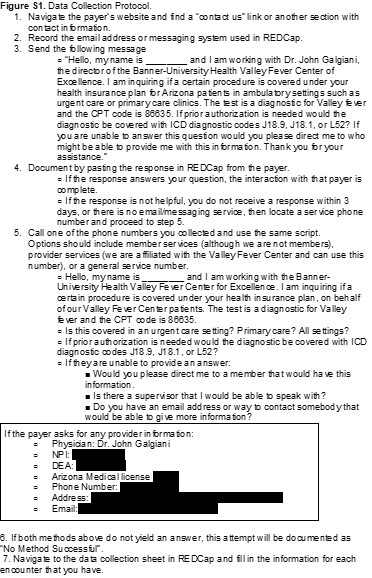
We used a predefined protocol to standardize the data collection process. When available, we contacted payers electronically through the use of built-in chat or messaging systems on the company websites, using a standardized message. We allowed 3 business days for a response. If they did not respond, did not provide an appropriate answer, or there was no messaging service available, we phoned the company using member or provider services. A copy of our data collection protocol is included in the supplemental materials (Figure S1).

Data collection and management used REDCap (Research Electronic Data Capture) hosted at The University of Arizona (version 11.3.4). REDCap is a secure, web-based software platform designed to support data capture for research studies (12,13). The list of variables obtained from communication with payer organizations included: payer name, method of communication used (message and telephone), department contacted, CPT 86635 coverage (including ICD-10 codes in the event of prior authorization), settings of coverage (urgent care, primary care, both), type of plan covered, and miscellaneous data including reason for non-disclosure of coverage. A copy of the complete data collection form is included in the supplemental materials (Figure S2).

Data Analysis

This is a descriptive study; no statistical significance testing was performed. Results are displayed as measures of frequency, including counts and percentages.

Results

Between 01/01/2021 and 09/21/2021 claims were submitted to 53 insurance payers. Duplicates and different plans under the same payer were merged and third-party claims processors were also excluded, resulting in 40 insurance payers for our study.

Data collection through contact with insurance companies occurred during September and October 2021. Of the 40 insurance payers identified, 12 (30.0%) had accessible online messaging via a messaging portal or email address. After messaging each of these payers with the scripted message, 6 responded. We contacted 35 (87.5%) insurance payers via telephone.

CPT 86635 was identified as covered in 15 (37.5%) of our communications (Figure 1).

Figure 1. Coverage of CPT code 86635 (Valley fever diagnostic tests) by 40 payers.

Of those 15 payers, 4 required prior authorization while 11 (73.3%) did not. All three of the ICD-10 codes (J18.1, J18.9, and L52) were accepted to obtain authorization. Those that did require prior authorization were either Department of Defense associated plans, or were not based out of Arizona, where coccidioidomycosis infections may not be as prominent.

Both the online message and phone script included differences in CPT coverage between an urgent care and primary care setting. Positive coverage responses that did not differentiate variations in coverage based on setting were recorded as covered in both urgent care and primary care. Of all positive coverages, 14 (93.3%) were covered in both urgent care and primary care and 1 (6.7%) did not specify if coverage was for both urgent care and primary care. Table 1 summarizes CPT coverage details.

Table 1. Coverage details for CPT code 86635 among payers (N = 40) a.

aList of payers (N=40) included: AARP Medicare, Aetna, All Savers, Allied Benefits System, Allwell, Ambetter, American Indian Health Program, ASR Health Benefits, AZ Foundation for Medical Care, Banner Family Care, BCBS Alabama, BCBS AZ, Bright Health, Care 1st Wellcare, CHAMP, Cigna, GEHA, Health Net, Humana, Imperial Health Texas Inc., Intel Arizona Connected Care, Kaiser Permanente, The Loomis Company, Medicare, Mercy Care, Meritain, Molina Complete Care of AZ, Multiplan Unified Life Insurance Company, OneCare Wellcare Medicare Advantage, Oscar Health Plan, Philadelphia American Life, Railroad MCR, Sierra Health and Life, Steward Health Choice, Summit, Tricare, Triwest VAPC, United Health, United Healthcare Community Plan, and WellCare MCR.

Of the 15 covered communications, 13 (86.7%) covered all plans, while 1 (6.7%) communication did not specify variation between plans, and 1 (6.7%) was member-specific to one of our researchers and denoted as “other”.

Of the 40 insurers contacted, 25 (62.5%) were unable to provide coverage information for Valley fever testing. The majority required member-specific information in order to disclose coverage details about a contracted plan. Given we had no specific patient for each plan and were only making general inquiries on behalf of a physician, we listed these communications as “Unable to Determine”. None of the 40 payers indicated that CPT code 86635 was not covered.

Discussion

In this study, we obtained coverage information for Valley fever diagnostic tests from 15 of the 40 payers we contacted. Of note, none of the remaining 25 payers said CPT code 86635 was not covered under their plans, only that they could not provide information, largely because such information requires specific member identification for one of their plan holders. In addition, 4 of the total providers required prior authorization for the diagnostic. These providers were either Department of Defense associated plans or were not based out of Arizona or California. Since coccidioidomycosis is largely endemic to Arizona and California, it is not unreasonable for an out of state insurance provider to require a prior authorization for a condition that is not endemic to their population. However, some national providers and out of state providers did state they cover the diagnostic without a prior authorization.

The difficulty of obtaining general coverage information from an insurance payer quickly became apparent. We anticipated that some payers would not disclose coverage information, however, given that we were requesting information on behalf of a practicing physician, we did not anticipate this response from most payers. The lack of transparency in providing benefit information to potential patients or providers is concerning and may negatively impact patient care. However, based upon the favorable response we received from payers that did provide information, it is likely that most of the insurers unable to provide information do cover the Valley fever diagnostic test.

Our findings build upon literature describing the lack of diagnoses of Valley fever, predominantly in the urgent care setting. Pu et al. (2020) reported the total diagnosis of coccidioidomycosis was a mere 0.5% in the urgent care setting from 2017-2019. At the time of our study, we found no previous publications on payer coverage of Valley fever diagnostic tests. However, we identified similar methods utilized in the existing literature. Cohen et al. (2019) researched insurance policies for coverage of gender re-affirming surgeries via online and telephonic methods and identified policies for 124 of 150 payers (14). A report by Park et al. (2019) researched insurance coverage policies for multiple pharmacogenomic tests via online methods and identified policies for 33 of 41 payers (15). Both of these studies were able to identify a larger proportion of coverage from the identified payers than our current study, though Park et al. (2019) did highlight difficulties from a patient or provider perspective in identifying payer coverage (13).

Results, however, must be considered in light of several study limitations. Payers were limited to those that were available via claims data from Sonora Quest Laboratories for predominantly Arizona payers. In addition, the claims data were derived solely from patients seen at Banner Health facilities, excluding patients seen for diagnosis and claims filed outside of the Banner Health network. The actual population of Valley fever patients is likely larger and may have had different coverage patterns than we collected. This data source and focus on Arizona limits generalizability of findings. However, Valley fever is endemic to Arizona and the Southwestern region of the United States.

This study also faced data collection limitations. Although our communications were scripted, the payers’ representatives may have not had a similar procedure. We may have obtained different results based upon the individual who was communicated with, and this may have impacted our ability to gather information.

For this study, we assumed that confirming CPT 86635 coverage by the payer’s representative meant coverage was generalizable to all plan types offered by the payer and all care settings where a patient might be seen. If a payer did not specify variability in coverage based on plan or care setting, we assumed all plans and all care settings were covered without need for prior authorization. 

Due to barriers that often exist for patients to see a primary care provider in a timely manner, many patients’ first interaction for Valley fever is in an urgent care setting. There remains a need to educate these providers about the availability and coverage of tests for patients, as current lack of knowledge may negatively impact patient care by delaying diagnoses and potentially leading to hospitalization.  While insurance coverage or cost may or may not be a limiting factor for a provider to order the diagnostic test, cost could be a limiting factor for the patient. Education can be provided to providers about recognition and testing coverage for Valley fever. Patients could then be educated as well in recognition of symptoms and insurance coverage trends, which could increase total tests ordered. Increased testing rates could help identify Valley fever diagnoses sooner and more frequently. This study highlights an important step of identifying payer coverage for Valley fever diagnosis in an urgent care setting. These results may help to inform providers about insurance coverage for their patients and increase early diagnosis of Valley fever cases. Future research could build upon this study by incorporating provider knowledge and education in relation to the impact on patients presenting with Valley fever in urgent care and primary care settings.

Acknowledgments

The authors wish to thank Banner Health and Sonora Quest Laboratories for their contributions in providing claims information for this research.

Author Contributions Statement

BVH, CEGR, JEK, and JAW contributed to data collection. CEGR, JEK, and JAW drafted the manuscript. AJG, JTH and JNG provided edits and commentary on the manuscript. All authors contributed to research design.

References

  1. Valley Fever Awareness. Centers for Disease Control and Prevention. Updated July 26, 2021. Accessed 12 October 2021. https://www.cdc.gov/fungal/features/valley-fever.html
  2. Galgiani JN. Coccidioidomycosis (coccidioides species). In: Bennett JE, Dolin R, Blaser MJ, eds. Mandell, Douglas, and Bennett's Principles and Practice of Infectious Diseases, 9th Ed. Philadelphia, PA: Elsevier; 2019.
  3. Valley Fever Statistics. Centers for Disease Control and Prevention. Accessed 12 October 2021. https://www.cdc.gov/fungal/diseases/coccidioidomycosis/statistics.html
  4. Kim MM, Blair JE, Carey EJ, Wu Q, Smilack JD. Coccidioidal pneumonia, Phoenix, Arizona, USA, 2000-2004. Emerg Infect Dis. 2009;15(3):397-401. doi:10.3201/eid1563.081007
  5. Valdivia L, Nix D, Wright M, et al. Coccidioidomycosis as a common cause of community-acquired pneumonia [published correction appears in Emerg Infect Dis. 2006 Aug;12(8):1307]. Emerg Infect Dis. 2006;12(6):958-962. doi:10.3201/eid1206.060028
  6. Donovan FM, Wightman P, Zong Y, et al. Delays in Coccidioidomycosis Diagnosis and Associated Healthcare Utilization, Tucson, Arizona, USA. Emerging Infectious Diseases. 2019;25(9):1745-1747. doi:10.3201/eid2509.190023.
  7. Pu J, Donovan FM, Ellingson K, et al. Clinician Practice Patterns That Result in the Diagnosis of Coccidioidomycosis Before or During Hospitalization. Clin Infect Dis. 2020;73(7):e1587-e1593. doi:10.1093/cid/ciaa739
  8. Grizzle AJ, Wilson L, Nix DE, Galgiani JN. Clinical and Economic Burden of Valley Fever in Arizona: An Incidence-Based Cost-of-Illness Analysis. Open Forum Infect Dis. 2020;8(2):ofaa623. Published 2020 Dec 28. doi:10.1093/ofid/ofaa623
  9. Learn More about Who We Are. Sonora Quest Laboratories. Accessed 15 October 2021. https://www.sonoraquest.com/about/who-we-are/.
  10. Galgiani JN, Ampel NM, Blair JE, et al. 2016 Infectious Diseases Society of America (IDSA) Clinical Practice Guideline for the Treatment of Coccidioidomycosis. Clin Infect Dis. 2016;63(6):e112-e146. doi:10.1093/cid/ciw360
  11. Donovan FM, Ramadan FA, Khan SA, et al. Comparison of a Novel Rapid Lateral Flow Assay to Enzyme Immunoassay Results for Early Diagnosis of Coccidioidomycosis. Clin Infect Dis. 2021;73(9):e2746-e2753. doi:10.1093/cid/ciaa1205
  12. Harris PA, Taylor R, Thielke R, Payne J, Gonzalez N, Conde JG. Research electronic data capture (REDCap)--a metadata-driven methodology and workflow process for providing translational research informatics support. J Biomed Inform. 2009;42(2):377-381. doi:10.1016/j.jbi.2008.08.010
  13. Harris PA, Taylor R, Minor BL, et al. The REDCap consortium: Building an international community of software platform partners. J Biomed Inform. 2019;95:103208. doi:10.1016/j.jbi.2019.103208
  14. Cohen WA, Sangalang AM, Dalena MM, Ayyala HS, Keith JD. Navigating Insurance Policies in the United States for Gender-affirming Surgery. Plast Reconstr Surg Glob Open. 2019 Dec 11;7(12):e2564. doi: 10.1097/GOX.0000000000002564. PMID: 32537307; PMCID: PMC7288898.
  15. Park SK, Thigpen J, Lee IJ. Coverage of pharmacogenetic tests by private health insurance companies. J Am Pharm Assoc (2003). 2020 Mar-Apr;60(2):352-356.e3. doi: 10.1016/j.japh.2019.10.003. Epub 2019 Dec 13. PMID: 31843376.

Cite as: Grace Rose CE, Kessler JE, Weisbrod JA, Hoang BV, Grizzle AJ, Hurwitz JT, Galgiani JN. Payer Coverage of Valley Fever Diagnostic Tests. Southwest J Pulm Crit Care. 2021;23(6):155-61. doi: https://doi.org/10.13175/swjpcc052-21 PDF